File:Kollmann425.jpg
Kollmann425.jpg (729 × 493 pixels, file size: 46 KB, MIME type: image/jpeg)
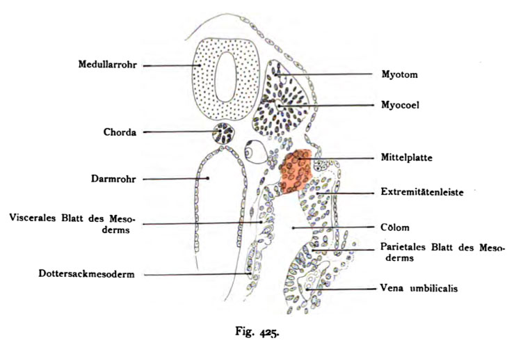
Fig. 425. Middle plate (intermediate mesoderm) human embryo of 13 somites, and 2.4 mm CRL
It is the left half of the cross-section.
(Anatomical Collection in Basel.)
Up next to the neural tube, the myotome is a myotome hole (Myocoel), the separation from the rest is almost complete ends, nor lie down cells, which are the central plate = inter-strand. connect The center plate is marked by red color. From this region of the embryonic body is formed, the pronephros, the Wolffian and See Müller course, even the inner part of the excretory system.
- This text is a Google translate computer generated translation and may contain many errors.
Images from - Atlas of the Development of Man (Volume 2)
(Handatlas der entwicklungsgeschichte des menschen)
- Kollmann Atlas 2: Gastrointestinal | Respiratory | Urogenital | Cardiovascular | Neural | Integumentary | Smell | Vision | Hearing | Kollmann Atlas 1 | Kollmann Atlas 2 | Julius Kollmann
- Links: Julius Kollman | Atlas Vol.1 | Atlas Vol.2 | Embryology History
| Historic Disclaimer - information about historic embryology pages |
|---|
 |
Reference
Kollmann JKE. Atlas of the Development of Man (Handatlas der entwicklungsgeschichte des menschen). (1907) Vol.1 and Vol. 2. Jena, Gustav Fischer. (1898).
Cite this page: Hill, M.A. (2024, April 27) Embryology Kollmann425.jpg. Retrieved from https://embryology.med.unsw.edu.au/embryology/index.php/File:Kollmann425.jpg
- © Dr Mark Hill 2024, UNSW Embryology ISBN: 978 0 7334 2609 4 - UNSW CRICOS Provider Code No. 00098G
Fig. 425. Mittelplatte (Zwischenstrans) eines tneoschlicheo Embryo
von 13 Urwirbeln und 2,4 mm gerader Länge. Es ist die linke Hälfte des Quer- schnittes dargestellt.
(Anatomische Sammlung in Basel.)
Oben neben dem Medullarrohr befindet sich das Myotom mit einer Myotom- höhle (Myocoel) ; die Trennung von dem übrigen Urwirbelgebiet ist nahezu vollendet, unten liegen noch Zellen, welche sich der Mittelplatte = Zwischenstrang anschließen. Die Mittelplatte ist durch rote Farbe ausgezeichnet. Aus diesem Gebiet des embryonalen Körpers entsteht die Vomiere, die Wolffschen und Müller sehen Gänge, überhaupt der innere Teil des exkretorischen Apparates.
File history
Click on a date/time to view the file as it appeared at that time.
| Date/Time | Thumbnail | Dimensions | User | Comment | |
|---|---|---|---|---|---|
| current | 14:23, 16 October 2011 | | 729 × 493 (46 KB) | S8600021 (talk | contribs) | {{Kollmann1907}} |
You cannot overwrite this file.
File usage
The following 2 pages use this file: