File:Keibel Mall 2 619.jpg
Original file (1,280 × 1,425 pixels, file size: 369 KB, MIME type: image/jpeg)
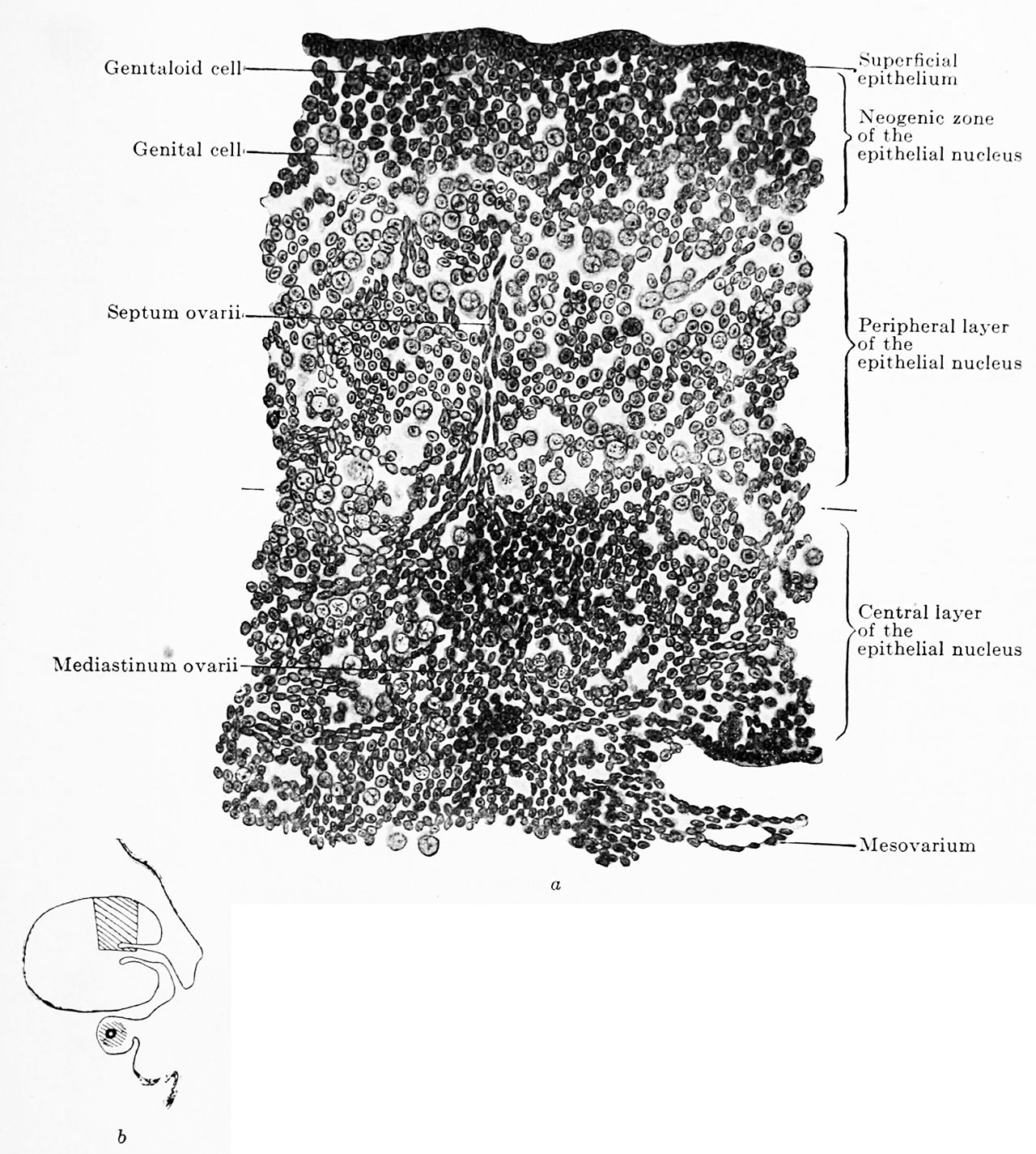
Fig. 619 a and b. Transverse section of the ovary of a human embryo of 80 mm trunk length, 100 mm head-foot length
(Embryo R. Meyer 151, from the collection of Professor R. Meyer, Berlin.) x230.
In Fig. 619 b the portion drawn is shown in the entire transverse section. The ovary has divided into the superficial epithelium, the neogenic zone, the peripheral and the central portions of the epithelial nucleus. Around the rete ovarii a kind of mediastinum has formed, from which a septum ovarii radiates towards the periphery. The neogenic zone consists of indifferent, genitaloid and genital cells, closely packed. The peripheral zone of the epithelial nucleus is composed of the same kinds of colls as the neogenic zone, but the cells are much more loosely arranged. In the rete blastema there are b still genitaloid cells.
| Embryology - 4 May 2024 |
|---|
| Google Translate - select your language from the list shown below (this will open a new external page) |
|
العربية | català | 中文 | 中國傳統的 | français | Deutsche | עִברִית | हिंदी | bahasa Indonesia | italiano | 日本語 | 한국어 | မြန်မာ | Pilipino | Polskie | português | ਪੰਜਾਬੀ ਦੇ | Română | русский | Español | Swahili | Svensk | ไทย | Türkçe | اردو | ייִדיש | Tiếng Việt These external translations are automated and may not be accurate. (More? About Translations) |
Felix W. The development of the urinogenital organs. In Keibel F. and Mall FP. Manual of Human Embryology II. (1912) J. B. Lippincott Company, Philadelphia. pp 752-979.
| Historic Disclaimer - information about historic embryology pages |
|---|
 |
Cite this page: Hill, M.A. (2024, May 4) Embryology Keibel Mall 2 619.jpg. Retrieved from https://embryology.med.unsw.edu.au/embryology/index.php/File:Keibel_Mall_2_619.jpg
- © Dr Mark Hill 2024, UNSW Embryology ISBN: 978 0 7334 2609 4 - UNSW CRICOS Provider Code No. 00098G
File history
Click on a date/time to view the file as it appeared at that time.
| Date/Time | Thumbnail | Dimensions | User | Comment | |
|---|---|---|---|---|---|
| current | 12:10, 12 November 2018 | | 1,280 × 1,425 (369 KB) | Z8600021 (talk | contribs) | |
| 12:08, 12 November 2018 | 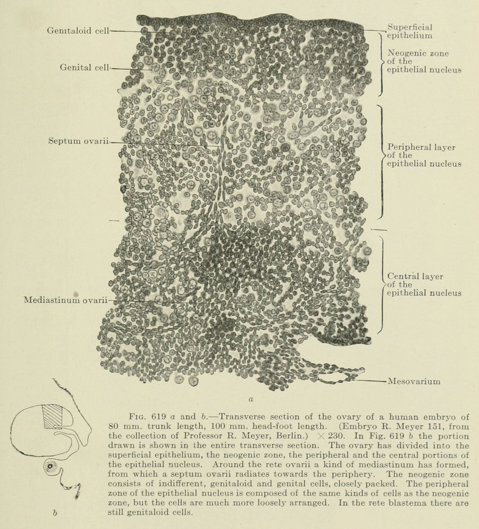 | 1,947 × 2,150 (627 KB) | Z8600021 (talk | contribs) |
You cannot overwrite this file.
File usage
The following 2 pages use this file: