File:Keibel Mall 2 610.jpg
Keibel_Mall_2_610.jpg (800 × 370 pixels, file size: 50 KB, MIME type: image/jpeg)
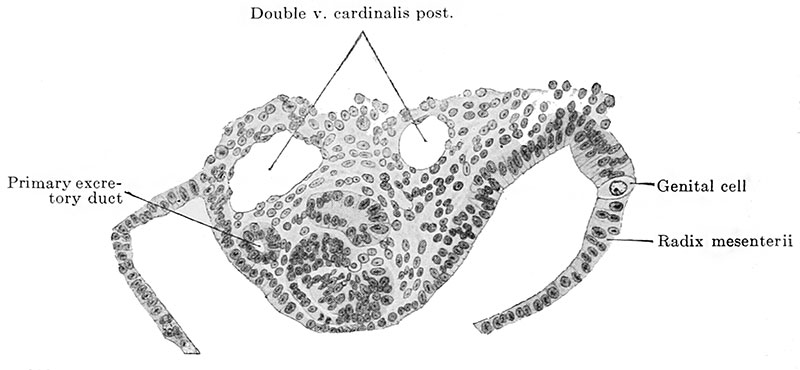
Fig. 610. Transverse section through an embryo of 4.7 mm vertex-breech length 33-35 pairs of primitive segments
Embryo of 4.7 mm vertex-breech length and 4.9 mm. nape length and with 33-35 pairs of primitive segments(Embryo 137, G. 31, from the collection of the II Anatomical Institute, Berlin, Professor O. Hertwig; slide 9, row 3, section 1.)
The section passes through the 11th primitive segment and the 5th mesonephric tubule. The urogenital fold is shown passing on the right into the root of the mesentery, on the left into the lateral wall of the body. A primary genital cell is to be seen in the root of the mesentery.
| Embryology - 4 May 2024 |
|---|
| Google Translate - select your language from the list shown below (this will open a new external page) |
|
العربية | català | 中文 | 中國傳統的 | français | Deutsche | עִברִית | हिंदी | bahasa Indonesia | italiano | 日本語 | 한국어 | မြန်မာ | Pilipino | Polskie | português | ਪੰਜਾਬੀ ਦੇ | Română | русский | Español | Swahili | Svensk | ไทย | Türkçe | اردو | ייִדיש | Tiếng Việt These external translations are automated and may not be accurate. (More? About Translations) |
Felix W. The development of the urinogenital organs. In Keibel F. and Mall FP. Manual of Human Embryology II. (1912) J. B. Lippincott Company, Philadelphia. pp 752-979.
| Historic Disclaimer - information about historic embryology pages |
|---|
 |
Cite this page: Hill, M.A. (2024, May 4) Embryology Keibel Mall 2 610.jpg. Retrieved from https://embryology.med.unsw.edu.au/embryology/index.php/File:Keibel_Mall_2_610.jpg
- © Dr Mark Hill 2024, UNSW Embryology ISBN: 978 0 7334 2609 4 - UNSW CRICOS Provider Code No. 00098G
File history
Click on a date/time to view the file as it appeared at that time.
| Date/Time | Thumbnail | Dimensions | User | Comment | |
|---|---|---|---|---|---|
| current | 08:44, 20 February 2014 | | 800 × 370 (50 KB) | Z8600021 (talk | contribs) | ==Fig. 610. Transverse section through an embryo of 4.7 mm. vertex-breech length and 4.9 mm. nape length and with 33-35 pairs of primitive segments== (Embryo 137, G. 31, from the collection of the II Anatomical Institute, Berlin, Professor O. Hertwig;... |
You cannot overwrite this file.
File usage
The following 2 pages use this file: