File:Keibel Mall 2 494.jpg
Original file (1,200 × 856 pixels, file size: 203 KB, MIME type: image/jpeg)
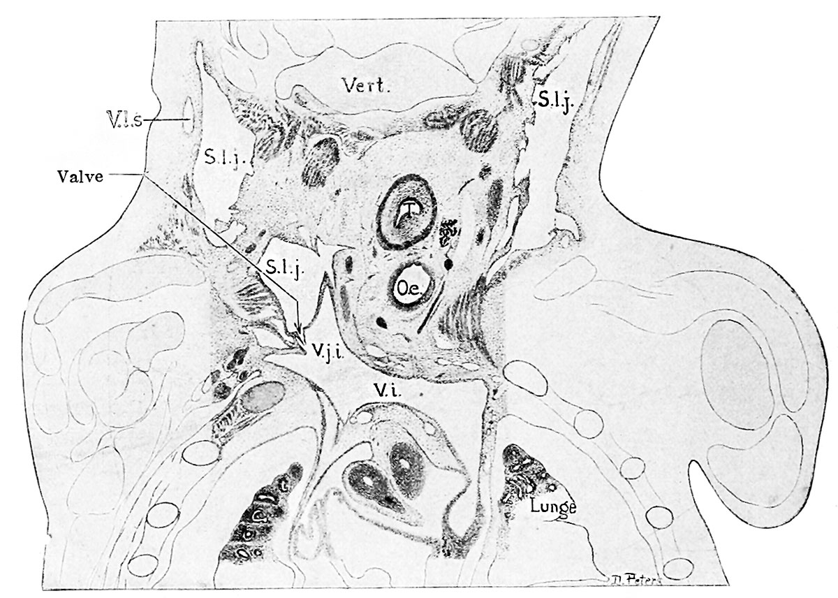
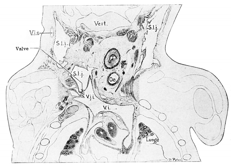
Fig. 494. Frontal section through the jugular lymph-sacs in a human embryo of 30 mm
Mall's collection, No. 86. x about 9.
The level of the section is shown on the reconstruction of Fig. 493. The section shows the complete lymph-sac on the right side and the valve on the left.
S.l.j., saccus lymphaticus jugularis; VS., v. innominata; V.j.i., v. jugularis interna; V.l.s., vasa lymphatica superficialis; Oe. , oesophagus; T., trachea.
- IV. The Development of the Lymphatic System: Chapter XVIII. Development of Blood, Vascular System, and Spleen | Historic Disclaimer
Reference
Sabin FR. The Development of the Lymphatic System in Keibel F. and Mall FP. Manual of Human Embryology II. (1912) J. B. Lippincott Company, Philadelphia.
Cite this page: Hill, M.A. (2024, May 27) Embryology Keibel Mall 2 494.jpg. Retrieved from https://embryology.med.unsw.edu.au/embryology/index.php/File:Keibel_Mall_2_494.jpg
- © Dr Mark Hill 2024, UNSW Embryology ISBN: 978 0 7334 2609 4 - UNSW CRICOS Provider Code No. 00098G
File history
Click on a date/time to view the file as it appeared at that time.
| Date/Time | Thumbnail | Dimensions | User | Comment | |
|---|---|---|---|---|---|
| current | 14:29, 2 March 2014 | | 1,200 × 856 (203 KB) | Z8600021 (talk | contribs) |
You cannot overwrite this file.
File usage
The following 2 pages use this file: