File:Keibel Mall 007.jpg
Original file (592 × 834 pixels, file size: 152 KB, MIME type: image/jpeg)
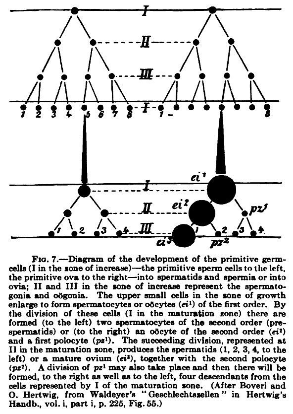
Fig. 7. Diagram of the development of the primitive germ cells
Diagram of the development of the primitive germ cells (I in the zone of increase) — the primitive sperm cells to the left, the primitive ova to the right — into spermatids and spermia or into ovia; II and III in the lone of increase represent the spermatogonia and odgonia. The upper small cells in the lone of growth enlarge to form spermatocytes or oocytes (etO of the first order. By the division of these cells (I in the maturation sone) there are formed (to the left) two spermatocytes of the second order (prespermatids) or (to the right) an oocyte of the second order (ei*) and a first olocyte (psO). The sucoeeding division, represented at II in the maturation sone, produces the spermatids (1, 2, 3, 4, to the left) or a mature ovum (et'), together with the second polocyte (pt*). A division of pz^ may also take place and then there will be formed, to the right as well as to the left, four descendants from the cells represented by I of the maturation zone.
(After Boveri and O. Hertwig, from Waldeyrer's "Geschlechtssellen" in Hertwig's Handb., vol. i, part i, p. 225, Fig. 55.
- KM Figure Links: The Germ Cells | Segmentation | First Primitive Segment | Gastrulation | External Form | Placenta | Axial Skeleton | Limb Skeleton | Skull | Muscular System
| Historic Disclaimer - information about historic embryology pages |
|---|
 |
Glossary Links
- Glossary: A | B | C | D | E | F | G | H | I | J | K | L | M | N | O | P | Q | R | S | T | U | V | W | X | Y | Z | Numbers | Symbols | Term Link
Cite this page: Hill, M.A. (2024, May 19) Embryology Keibel Mall 007.jpg. Retrieved from https://embryology.med.unsw.edu.au/embryology/index.php/File:Keibel_Mall_007.jpg
- © Dr Mark Hill 2024, UNSW Embryology ISBN: 978 0 7334 2609 4 - UNSW CRICOS Provider Code No. 00098G
File history
Click on a date/time to view the file as it appeared at that time.
| Date/Time | Thumbnail | Dimensions | User | Comment | |
|---|---|---|---|---|---|
| current | 20:10, 12 February 2012 | | 592 × 834 (152 KB) | S8600021 (talk | contribs) | ==Fig. 7. == {{Keibel_Mall Images}} |
You cannot overwrite this file.
File usage
The following 2 pages use this file: