File:Keibel1910 fig13.jpg
Keibel1910_fig13.jpg (520 × 520 pixels, file size: 25 KB, MIME type: image/jpeg)
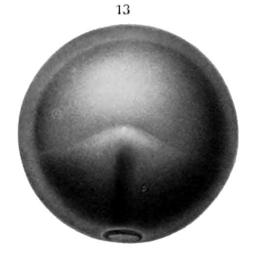
Fig. 13. Top view of egg 13 days 3 hrs old
Top view of egg 13 days 3 hrs. old. Small circular blastopore. Embryonic anläge triangulär in outline ; lateral boundaries indistinct. First appearance of neural groove. Roof of segmentation cavity thinner, making its boundaries distinct. (X 10.)
| Historic Disclaimer - information about historic embryology pages |
|---|
 |
Reference
Eycleshymer AC. and Wilson JM. Normal Plates of the Development of the Salamander Embryo (Nectürüs maculosus). Vol. 11 in series by Keibel F. Normal plates of the development of vertebrates (Normentafeln zur Entwicklungsgeschichte der Wirbelthiere) Fisher, Jena., Germany.
Cite this page: Hill, M.A. (2024, May 21) Embryology Keibel1910 fig13.jpg. Retrieved from https://embryology.med.unsw.edu.au/embryology/index.php/File:Keibel1910_fig13.jpg
- © Dr Mark Hill 2024, UNSW Embryology ISBN: 978 0 7334 2609 4 - UNSW CRICOS Provider Code No. 00098G
File history
Click on a date/time to view the file as it appeared at that time.
| Date/Time | Thumbnail | Dimensions | User | Comment | |
|---|---|---|---|---|---|
| current | 14:04, 10 January 2015 | | 520 × 520 (25 KB) | Z8600021 (talk | contribs) | ==Fig. 13. Top view of egg 13 days 3 hrs old== Top view of egg 13 days 3 hrs. old. Small circular blastopore. Embryonic anläge triangulär in outline ; lateral boundaries indistinct. First appearance of neural groove. Roof of segmentation cavity thin... |
You cannot overwrite this file.
File usage
The following 3 pages use this file: