File:Keibel1908 plate01.jpg
From Embryology

Size of this preview: 434 × 600 pixels. Other resolution: 1,447 × 2,000 pixels.
Original file (1,447 × 2,000 pixels, file size: 437 KB, MIME type: image/jpeg)
Plate 1.
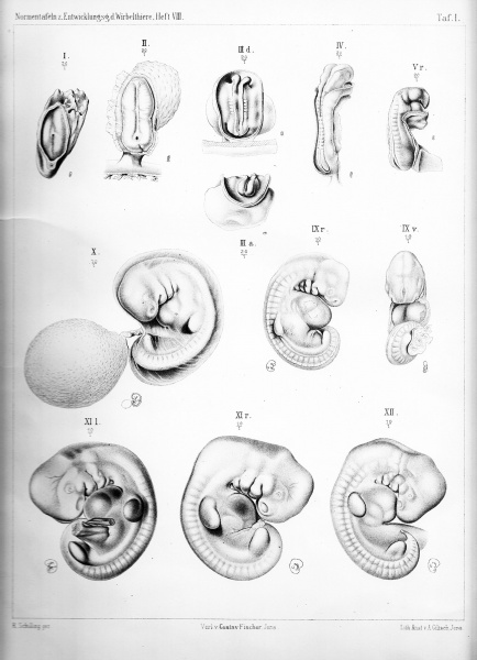
- The design of the actual embryonic body begins with a stage, as shown him in the standard panel Figure I shows us. In a relatively large yolk sac lies flat spread the germ shield with primitive streak and neurenteric canal, in front of the neurenteric canal a shallow medullary groove expands, which is flanked by shallow, not sharply defined medulla bulges. (p84)
- The emergence of the brain part relative to the spinal cord part of the central nervous system makes early claims. Even in an embryo, 6-7 mesoblastic somite pairs (standard panel Figure III) are the three primary brain divisions are distinguishable and the cephalic flexure has occurred. (p84)
- In the Figs. IV and V of the standard panel the cranial and caudal medullary tube is still open.
- In the Figs. IV and V of the standard panel the cranial and caudal medullary tube is still open.
- The neural tube closes initially at the cranial end; on the embryo which is shown in Figure VI of the standard panel, the closing position of the front neuropore can be seen more precisely; (p84)
- In the embryo of Figure VII the posterior neuropore very close to the end. The caudal end of the medullary system does not result from the formation and closure of medullary fold, but differentiates itself with notochord and tail gut can call from the indifferent cell mass, which we see arise at the caudal embryonic end after shrinkage of the primitive streak and the tail bud. (p85)
- The peak of the curvature is seen in the embryos of Figs. X and XI of the standard panel. Now the body begins to stretch, perhaps under the influence of himself more and more from the forming liver, which is also reflected in the surface image under the cardiac prominence more and more claims and this probably holds the balance in Figure XIV. (p86)
- Vol. 8 Human: Fig 1 | Fig 2 | Fig 3 | Fig 4 | Fig 5 | Fig 6 | Fig 7 | Fig 8 | Fig 9 | Fig 10 | Fig 11 | Fig 12 | Fig 13 | Fig 14 | Fig 15 | Fig 16a | Fig 16b | Fig 17 | Fig 18 | Fig 19 | Fig 20 | Fig 21 | Fig 22 | Fig 23 | Fig 24 | Fig 25 | Fig 27 | Fig 27 | Fig 28 | Fig 29 | Fig 30a | Fig 30b | Fig 31 | Fig 32 | Fig 33 | Fig 34a | Fig 34b | Fig 35 | Fig 36 | Fig 37 | Fig 38 | Fig 39 | Fig 40 | Fig 41 | Fig 42 | Fig 43 | Fig 44 | Plate 1 | Plate 2 | Plate 3 | Plate 4 | Plate 5 | Plate 6 | Franz Keibel | Embryonic Development
| Historic Disclaimer - information about historic embryology pages |
|---|
 |
Reference
Keibel F. and Elze C. Normal Plates of the Development of the Human Embryo (Homo sapiens). (1908) Vol. 8 in series by Keibel F. Normal plates of the development of vertebrates (Normentafeln zur Entwicklungsgeschichte der Wirbelthiere) Fisher, Jena., Germany.
Cite this page: Hill, M.A. (2024, April 27) Embryology Keibel1908 plate01.jpg. Retrieved from https://embryology.med.unsw.edu.au/embryology/index.php/File:Keibel1908_plate01.jpg
- © Dr Mark Hill 2024, UNSW Embryology ISBN: 978 0 7334 2609 4 - UNSW CRICOS Provider Code No. 00098G
File history
Click on a date/time to view the file as it appeared at that time.
| Date/Time | Thumbnail | Dimensions | User | Comment | |
|---|---|---|---|---|---|
| current | 23:38, 30 November 2013 | | 1,447 × 2,000 (437 KB) | Z8600021 (talk | contribs) | |
| 23:37, 30 November 2013 |  | 1,447 × 2,000 (589 KB) | Z8600021 (talk | contribs) | ||
| 23:40, 17 November 2013 | 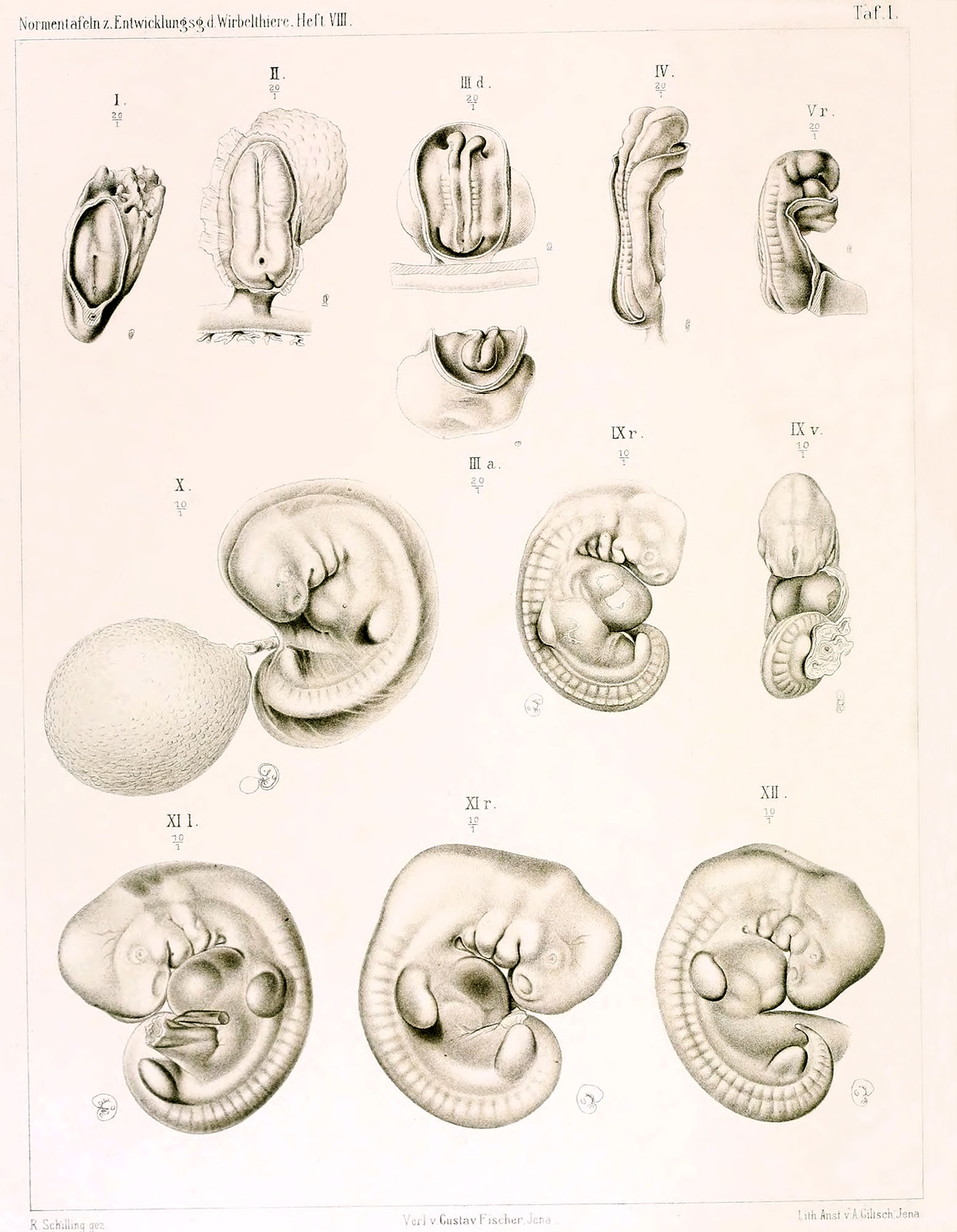 | 1,554 × 2,000 (370 KB) | Z8600021 (talk | contribs) | {{Keibel1908figures}} |
You cannot overwrite this file.
File usage
The following 11 pages use this file:
- Book - Normal plates of the development of the human embryo (1908)
- Carnegie stage table
- Development Group Meeting 2017 - Digital Embryology Consortium
- Embryology History - Franz Keibel
- Gottingen Meeting 2017 - Digital Embryology Consortium
- Lecture - 2014 Course Introduction
- Lecture - 2015 Course Introduction
- Lecture - 2016 Course Introduction
- Lecture - 2017 Course Introduction
- Talk:Berlin Meeting 2017 - Digital Embryology Consortium
- Talk:Lecture - 2016 Course Introduction