File:Ingalls1920plate04.jpg
Original file (762 × 1,044 pixels, file size: 52 KB, MIME type: image/jpeg)
Plate 4
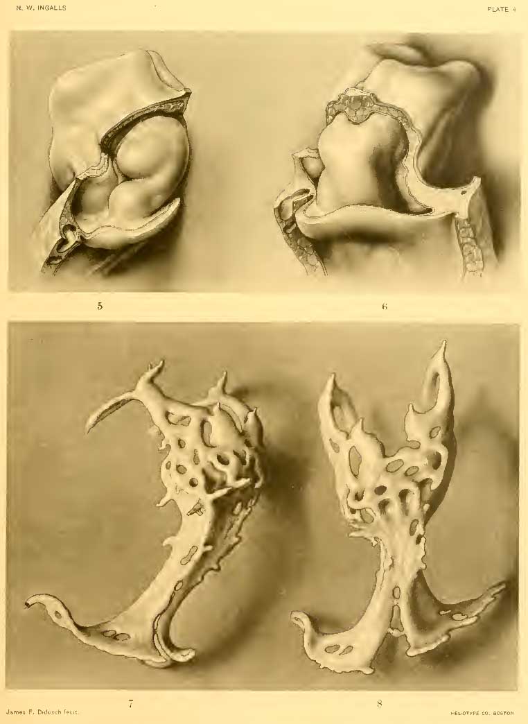
Fig. 5.
Right ventrolateral view of heart, myoepicardial mantle, and pericardial cavity. X 100.
Fig. 6.
Ventral wall, toward exocecelom, has been removed (cf. fig. 3). Ventral view of same model, slightly from the left side.
Fig. 7.
The vein in the splanchnopleure on the right side is the yolk-sac portion of the omphalomesenteric vein, as yet unconnected with the heart.
Fig. 8.
Right ventrolateral view of cardiac plexus. X 200. Same model in ventral view.
For position of heart plexus within the body, see figures 3 and 4.
- Embryo at Segmentation: Figure A | Plate 1 | Plate 2 | Plate 3 | Plate 4 | Plate 5 | Carnegie stage 9 | Carnegie Embryo 1878
Reference
Ingalls NW. A human embryo at the beginning of segmentation, with special reference to the vascular system. (1920) Contrib. Embryol., Carnegie Inst. Wash. Publ. 274, 11: 61-90.
Cite this page: Hill, M.A. (2024, April 28) Embryology Ingalls1920plate04.jpg. Retrieved from https://embryology.med.unsw.edu.au/embryology/index.php/File:Ingalls1920plate04.jpg
- © Dr Mark Hill 2024, UNSW Embryology ISBN: 978 0 7334 2609 4 - UNSW CRICOS Provider Code No. 00098G
| Historic Disclaimer - information about historic embryology pages |
|---|
 |
File history
Click on a date/time to view the file as it appeared at that time.
| Date/Time | Thumbnail | Dimensions | User | Comment | |
|---|---|---|---|---|---|
| current | 18:05, 30 January 2012 | | 762 × 1,044 (52 KB) | S8600021 (talk | contribs) | {{Ingalls1920}} {{Historic Disclaimer}} |
You cannot overwrite this file.
File usage
The following 4 pages use this file: