File:Ingalls1908 plate01.jpg
From Embryology
Size of this preview: 556 × 600 pixels. Other resolution: 927 × 1,000 pixels.
Original file (927 × 1,000 pixels, file size: 204 KB, MIME type: image/jpeg)
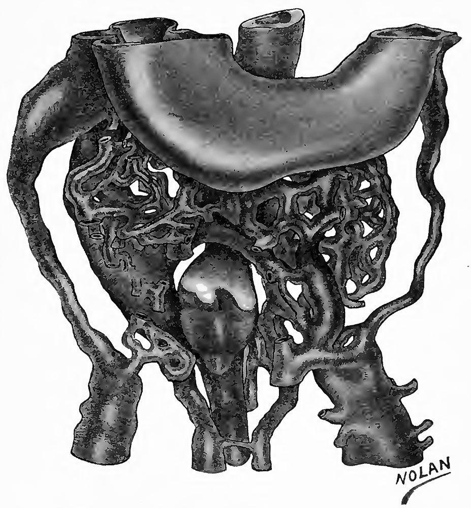
Plate I Model of veins, ventral view
The conneotion of the liver and gut has been severed just above the gall-bladder anlage. Just above this is seen the ventral half of the cranial venous ring in connection with both omphalo-mesenteric, and the left umbilical veins. X 100
| Historic Disclaimer - information about historic embryology pages |
|---|
 |
Reference
Ingalls NW. A contribution to the embryology of the liver and vascular system in man. (1908) Anat. Rec. 2: 338–344.
Cite this page: Hill, M.A. (2024, May 6) Embryology Ingalls1908 plate01.jpg. Retrieved from https://embryology.med.unsw.edu.au/embryology/index.php/File:Ingalls1908_plate01.jpg
- © Dr Mark Hill 2024, UNSW Embryology ISBN: 978 0 7334 2609 4 - UNSW CRICOS Provider Code No. 00098G
File history
Click on a date/time to view the file as it appeared at that time.
| Date/Time | Thumbnail | Dimensions | User | Comment | |
|---|---|---|---|---|---|
| current | 22:41, 15 February 2016 | | 927 × 1,000 (204 KB) | Z8600021 (talk | contribs) | |
| 22:40, 15 February 2016 | 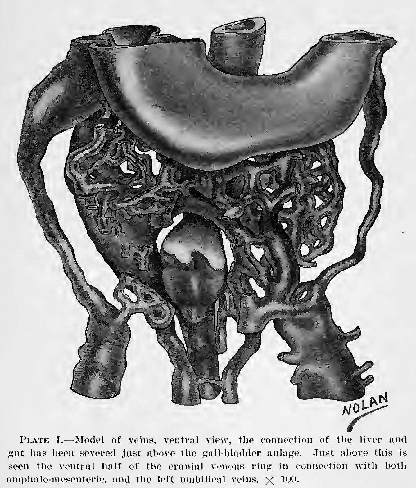 | 1,340 × 1,571 (423 KB) | Z8600021 (talk | contribs) |
You cannot overwrite this file.
File usage
The following page uses this file: