File:Hypothyroidism.jpg

Original file (1,492 × 1,634 pixels, file size: 565 KB, MIME type: image/jpeg)
Hyperthyroidism
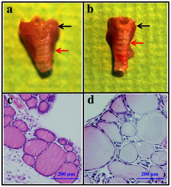
This image compares a normal thyroid gland (a) anatomy with a hypothyroidism thyroid gland (b). The histology images reveal in the abnormal thyroid gland, there is less thyroid hormone produced due to irregular epithelium on the surface of the follicles. The researchers in this report found that once thyroid stimulating hormone was added to the hypothyroid follicles, the epithelium became more columnar and the lumens became smaller, similar to healthy thyroid follicles.
Reference
<pubmed>22916127</pubmed>
Copyright © Endo, Kobayashi. This is an open-access article distributed under the terms of the Creative Commons Attribution License, which permits unrestricted use, distribution, and reproduction in any medium, provided the original author and source are credited.
- Note - This image was originally uploaded as part of an undergraduate science student project and may contain inaccuracies in either description or acknowledgements. Students have been advised in writing concerning the reuse of content and may accidentally have misunderstood the original terms of use. If image reuse on this non-commercial educational site infringes your existing copyright, please contact the site editor for immediate removal.
File history
Click on a date/time to view the file as it appeared at that time.
| Date/Time | Thumbnail | Dimensions | User | Comment | |
|---|---|---|---|---|---|
| current | 09:18, 24 October 2014 | | 1,492 × 1,634 (565 KB) | Z3414648 (talk | contribs) | This image compares a normal thyroid gland (a) anatomy with a hypothyroidism thyroid gland (b). The histology images reveal in the abnormal thyroid gland, there is less thyroid hormone produced due to irregular epithelium on the surface of the follicle... |
You cannot overwrite this file.
File usage
The following 2 pages use this file: