File:Human stage14 heart MRI - ventricular septation.jpg
From Embryology

Size of this preview: 795 × 600 pixels. Other resolution: 864 × 652 pixels.
Original file (864 × 652 pixels, file size: 39 KB, MIME type: image/jpeg)
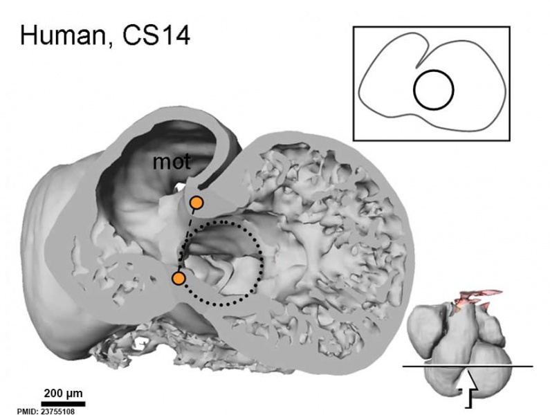
Human Heart Ventricular Septation (Carnegie stage 14)
Miniature show sectioning plane and angle of inspection.
- orange dot - bulbo-ventricular fold on the border of the trabeculated ventricle and the myocardial outflow tract.
- circle - The atrioventricular canal is exclusively to the left of the fold.
- broken line - ventricular septation
- Links: Image - stage 14 | Carnegie stage 14 | Image - stage 18 | Carnegie stage 18 | Cardiovascular System Development | Magnetic Resonance Imaging
Reference
<pubmed>23755108</pubmed>| PLoS One.
Copyright
© 2013 Jensen et al. This is an open-access article distributed under the terms of the Creative Commons Attribution License, which permits unrestricted use, distribution, and reproduction in any medium, provided the original author and source are credited.
Figure 11. Comparative ventricular development. doi:10.1371/journal.pone.0063651.g011 Human heart panel cropped, resized and relabelled.
File history
Click on a date/time to view the file as it appeared at that time.
| Date/Time | Thumbnail | Dimensions | User | Comment | |
|---|---|---|---|---|---|
| current | 14:28, 13 September 2014 | | 864 × 652 (39 KB) | Z8600021 (talk | contribs) | |
| 14:25, 13 September 2014 | 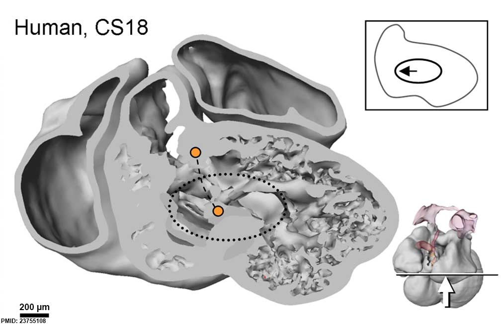 | 1,014 × 652 (48 KB) | Z8600021 (talk | contribs) |
You cannot overwrite this file.
File usage
The following page uses this file: