File:Human embryo day 18.jpg
Original file (531 × 800 pixels, file size: 80 KB, MIME type: image/jpeg)
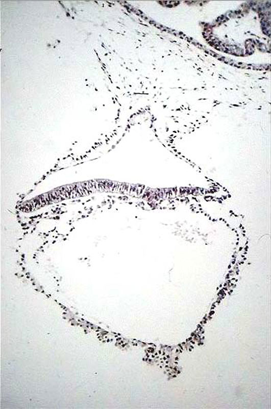
Human Embryo (day 18)
Approximate cross-section of a different, 18 day human conceptus (Carnegie stage 6).
Identify the
- 3 cavities - yolk sac, amniotic sac and chorionic sac.
- 3 layers of the trilaminar embryo - ectoderm (columnar cells), intraembryonic mesoderm (mesenchymal cells, endodermal cells (cuboidal single layer).
- extraembryonic mesoderm which surrounds the yolk and amniotic sacs, and continues to form the connecting stalk, where it then continues to line the chorionic sac. Extra- and intra-embryonic mesoderm merge along the perimeter of the embryonic disc.
- primitive groove with dense cluster of primitive streak cells below it.
At the top right of the micrograph is a secondary chorionic villus, lying in an empty maternal intervillus space. The villous has an outer layer of syncytiotrophoblast (smaller, darker nuclei), an intermediate layer of cytotrophoblast and a core of mesoblast (extra-embryonic mesoderm).
- Links: Carnegie stage 6
Reference
Nishimura etal., 1977
Cite this page: Hill, M.A. (2024, May 11) Embryology Human embryo day 18.jpg. Retrieved from https://embryology.med.unsw.edu.au/embryology/index.php/File:Human_embryo_day_18.jpg
- © Dr Mark Hill 2024, UNSW Embryology ISBN: 978 0 7334 2609 4 - UNSW CRICOS Provider Code No. 00098G
File history
Click on a date/time to view the file as it appeared at that time.
| Date/Time | Thumbnail | Dimensions | User | Comment | |
|---|---|---|---|---|---|
| current | 21:48, 8 October 2015 | | 531 × 800 (80 KB) | Z8600021 (talk | contribs) | |
| 20:38, 9 May 2010 |  | 398 × 600 (38 KB) | S8600021 (talk | contribs) | Human embryo day 18 Category:Week 3 |
You cannot overwrite this file.
File usage
The following 7 pages use this file: