File:Hertig1946b fig12.jpg
Original file (800 × 1,333 pixels, file size: 229 KB, MIME type: image/jpeg)
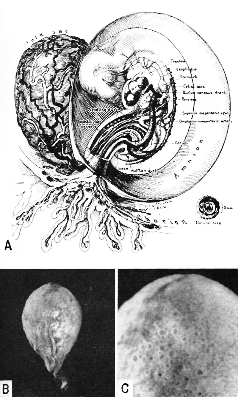
Fig. 12. Human Embryo 5 to 6.7 mm
A. A drawing of a 5 mm embryo (late 4th or early 5th week of development) showing further development of the umbilical cord. The yolk-sac is large, well vascularized and its wall contains numerous cystic and tubular spaces derived from the lining epithelium. (Fig. 5 from Cullen’s “The Umbilicus and Its Diseases,” W. B. Saunders Company.)
B. A photograph of the yolk-sac of a 6.7 mm. embryo. Carnegie 7522, sequence 9, X8.
C. A higher power photograph of the same yolksac as seen in Fig. B. to show the numerous cystic and gland-like spaces in its wall. Carnegie 7522, sequence 7, X22.
References
Hertig AT. lnvolution of tissues in fetal life: a review. (1946) Anat. Rec. 94: 96-116.
Cite this page: Hill, M.A. (2024, May 10) Embryology Hertig1946b fig12.jpg. Retrieved from https://embryology.med.unsw.edu.au/embryology/index.php/File:Hertig1946b_fig12.jpg
- © Dr Mark Hill 2024, UNSW Embryology ISBN: 978 0 7334 2609 4 - UNSW CRICOS Provider Code No. 00098G
File history
Click on a date/time to view the file as it appeared at that time.
| Date/Time | Thumbnail | Dimensions | User | Comment | |
|---|---|---|---|---|---|
| current | 08:42, 8 August 2017 | | 800 × 1,333 (229 KB) | Z8600021 (talk | contribs) | ===References=== {{Ref-Hertig1946b}} {{Footer}} Category:Carnegie Embryo 6344 |
You cannot overwrite this file.
File usage
The following page uses this file: