File:Heart valve histology 02.jpg
Heart_valve_histology_02.jpg (800 × 456 pixels, file size: 103 KB, MIME type: image/jpeg)
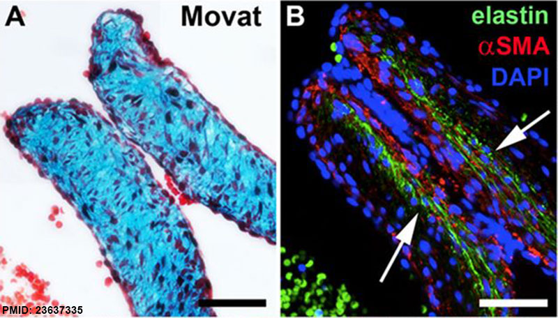
Developing Heart Valve Histology and Gene Expression
Human fetal (GA week 9) semilunar valve leaflet section.
- A - Movat's stain
- B - immunofluorescence staining with an antibody against tropoelastin/elastin (green) of DAPI-stained nuclei (blue) and α-smooth muscle actin (αSMA)-expressing cells (red).
Arrows indicate tropoelastin/elastin in the layer facing the ventricles.
Scale bars: 50 μm
Reference
<pubmed>23637335</pubmed>| Development
Copyright
© 2013. Published by The Company of Biologists Ltd This is an Open Access article distributed under the terms of the Creative Commons Attribution License (http://creativecommons.org/licenses/by/3.0), which permits unrestricted use, distribution and reproduction in any medium provided that the original work is properly attributed.
File history
Click on a date/time to view the file as it appeared at that time.
| Date/Time | Thumbnail | Dimensions | User | Comment | |
|---|---|---|---|---|---|
| current | 18:23, 4 March 2015 | | 800 × 456 (103 KB) | Z8600021 (talk | contribs) | ==Developing Heart Valve Histology and Gene Expression== Human fetal ({{GA}} week 9) semilunar valve leaflet section. * A - Movat's stain * B - immunofluorescence staining with an antibody against tropoelastin/elastin (green) of DAPI-stained nuclei (... |
You cannot overwrite this file.
File usage
There are no pages that use this file.