File:Heart Looping Sequence.jpg
From Embryology
Size of this preview: 800 × 298 pixels. Other resolution: 1,548 × 577 pixels.
Original file (1,548 × 577 pixels, file size: 85 KB, MIME type: image/jpeg)
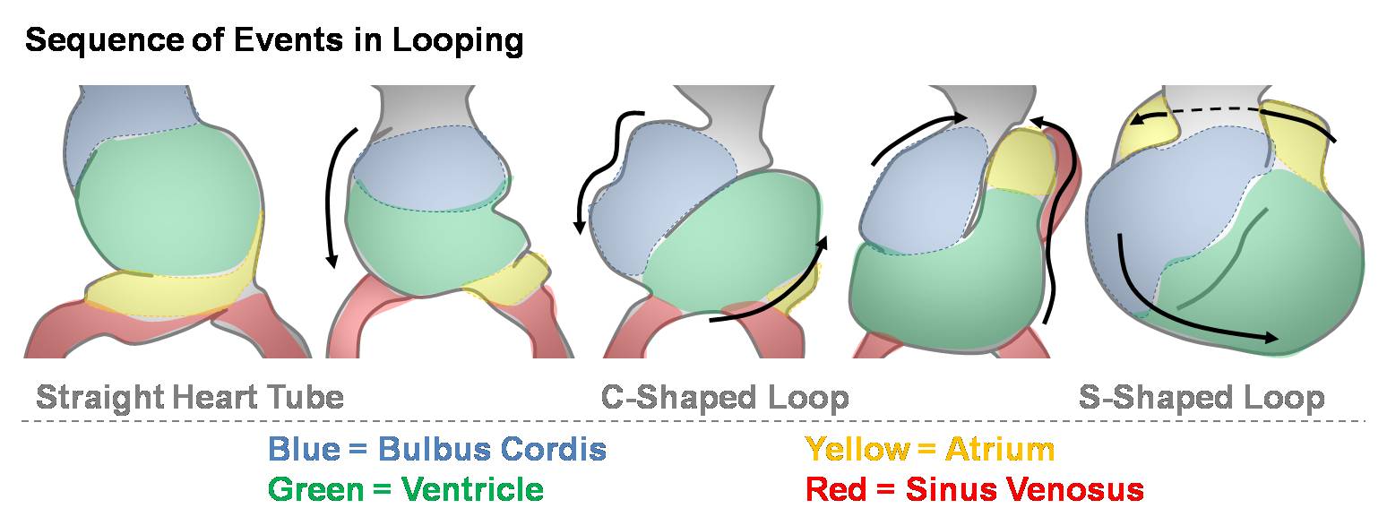
The Sequence of Events in Heart Looping
Week 4 to week 5 (GA 6 to 7 weeks)
- The heart begins as a straight tube then bends ventrally.
- Rotation brings the bulge of the ventral bend (predominantly the bulbus cordis and ventricle) to the right, forming a "C-shaped" loop (sometimes called "U-shaped").
- As the atrium is brought cranially, the poles of the heart converge. The heart now appears as an "S-shape", with the first bend in the S between the bulbus cordis and ventricle and the second bend between the atrium and sinus venosus.
- The bulbus cordis and arterial trunk move ventral to the atrium to form the later outflow tract. The atrium now lies superior to the ventricle.

| Begin Intermediate: | Primordial Heart Tube | Heart Tube Looping | Atrial Ventricular Septation | Outflow Tract | Heart Valves | Cardiac Abnormalities | Vascular Overview |
Cite this page: Hill, M.A. (2024, May 13) Embryology Heart Looping Sequence.jpg. Retrieved from https://embryology.med.unsw.edu.au/embryology/index.php/File:Heart_Looping_Sequence.jpg
- © Dr Mark Hill 2024, UNSW Embryology ISBN: 978 0 7334 2609 4 - UNSW CRICOS Provider Code No. 00098G
File history
Click on a date/time to view the file as it appeared at that time.
| Date/Time | Thumbnail | Dimensions | User | Comment | |
|---|---|---|---|---|---|
| current | 10:49, 14 March 2010 | 1,548 × 577 (85 KB) | Z3212774 (talk | contribs) | category:Heart ILP Shows the sequence of events in heart looping. The heart begins as a straight tube then bends ventrally. Rotation brings the bulge of the ventral bend (predominantly the bulbus cordis and ventricle) to the right, forming a C-shaped |
You cannot overwrite this file.
File usage
The following file is a duplicate of this file (more details):