File:Hamilton1949 fig10.jpg

Original file (1,250 × 1,003 pixels, file size: 162 KB, MIME type: image/jpeg)
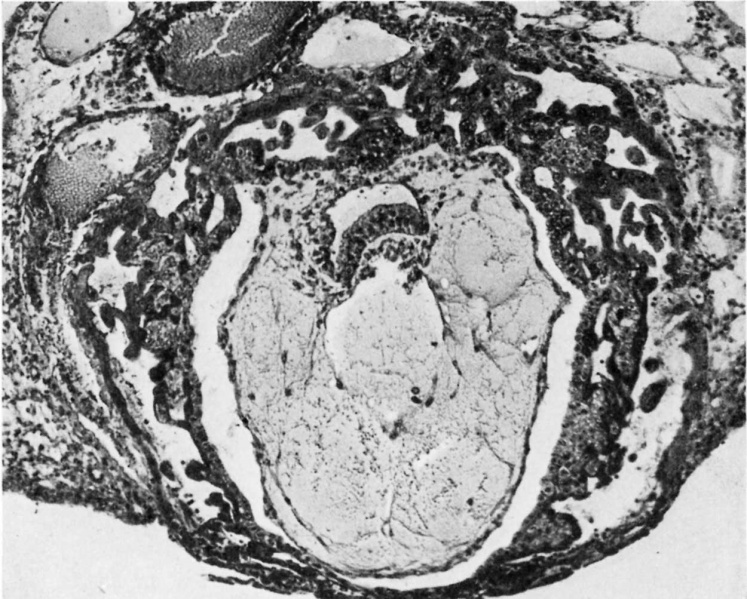
Fig.10. A general low-power view of the Barnes embryo
Estimated to be about 10.5 days old. The extent of the syncytio- and cyto-trophoblast in the different parts of the vesicle is shown. The embryonic rudiment has differentiated into columnar ectoderm and embryonic endoderm, the latter is continuous with Heuser's membrane. The extra-embryonic mesoblast has been artificialy separated from the cytotrophoblast. X.C.
Reference
Hamilton WJ. Early stages of human development. (1949) Ann R Coll Surg Engl. 4(5): 281-94. PMID 18121228 PMC2238331
Cite this page: Hill, M.A. (2024, May 4) Embryology Hamilton1949 fig10.jpg. Retrieved from https://embryology.med.unsw.edu.au/embryology/index.php/File:Hamilton1949_fig10.jpg
- © Dr Mark Hill 2024, UNSW Embryology ISBN: 978 0 7334 2609 4 - UNSW CRICOS Provider Code No. 00098G
File history
Click on a date/time to view the file as it appeared at that time.
| Date/Time | Thumbnail | Dimensions | User | Comment | |
|---|---|---|---|---|---|
| current | 15:38, 15 May 2018 | | 1,250 × 1,003 (162 KB) | Z8600021 (talk | contribs) | |
| 15:37, 15 May 2018 | 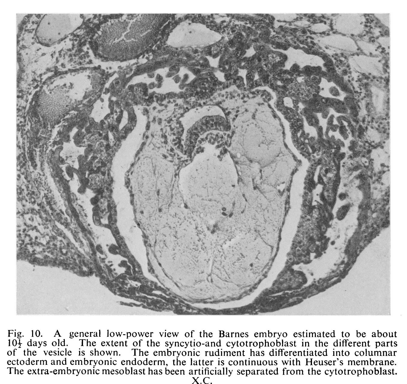 | 1,358 × 1,281 (304 KB) | Z8600021 (talk | contribs) |
You cannot overwrite this file.
File usage
The following page uses this file: