File:Gray1114.jpg
From Embryology
No higher resolution available.
Gray1114.jpg (450 × 471 pixels, file size: 47 KB, MIME type: image/jpeg)
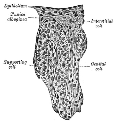
Human Embryo (3.5 cm long) Testis Section of a Genital Cord
(Felix and Bühler.)
| Historic Disclaimer - information about historic embryology pages |
|---|
 |
The Testis
- testis is developed in much the same way as the ovary
- earliest stages it consists of a central mass of epithelium covered by a surface epithelium
- in the central mass a series of cords appear
- periphery of the mass is converted into the tunica albuginea
- excluding the surface epithelium from any part in the formation of the tissue of the testis
- cords of the central mass run together toward the future hilus
- form a network which ultimately becomes the rete testis
- cords develop the seminiferous tubules
- between them connective-tissue septa extend
- seminiferous tubules become connected with outgrowths from the Wolffian body
- form the efferent ducts of the testis
(text modified from Gray's Anatomy)
- Gray's Images: Development | Lymphatic | Neural | Vision | Hearing | Somatosensory | Integumentary | Respiratory | Gastrointestinal | Urogenital | Endocrine | Surface Anatomy | iBook | Historic Disclaimer
| Historic Disclaimer - information about historic embryology pages |
|---|
|
| iBook - Gray's Embryology | |
|---|---|

|
|
Reference
Gray H. Anatomy of the human body. (1918) Philadelphia: Lea & Febiger.
Cite this page: Hill, M.A. (2024, April 30) Embryology Gray1114.jpg. Retrieved from https://embryology.med.unsw.edu.au/embryology/index.php/File:Gray1114.jpg
- © Dr Mark Hill 2024, UNSW Embryology ISBN: 978 0 7334 2609 4 - UNSW CRICOS Provider Code No. 00098G
File history
Click on a date/time to view the file as it appeared at that time.
| Date/Time | Thumbnail | Dimensions | User | Comment | |
|---|---|---|---|---|---|
| current | 08:44, 28 May 2011 | | 450 × 471 (47 KB) | S8600021 (talk | contribs) | ==Human Embryo (3.5 cm long) Testis Section of a Genital Cord== (Felix and Bühler.) {{Historic Disclaimer}} (text modified from Gray's Anatomy) {{Gray Anatomy}} Category:Human Category:Genital Category:Male Category:Testis |
You cannot overwrite this file.
File usage
The following 5 pages use this file: