File:Gray1109.jpg
From Embryology
No higher resolution available.
Gray1109.jpg (464 × 487 pixels, file size: 56 KB, MIME type: image/jpeg)
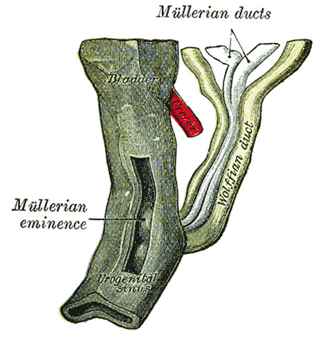
Fig. 1109. Urogenital Sinus of Female Human Embryo of 8.5 to 9 weeks old
(From model by Keibel)
| Historic Disclaimer - information about historic embryology pages |
|---|
 |
The Müllerian Ducts (Paramesonephric Ducts)
- Shortly after the formation of the Wolffian ducts a second pair of ducts is developed, the Müllerian ducts
- Each arises on the lateral aspect of the corresponding Wolffian duct as a tubular invagination of the cells lining the coelom
- orifice of the invagination remains patent, and undergoes enlargement and modification to form the abdominal ostium of the uterine tube
- ducts pass backward lateral to the Wolffian ducts
- toward the posterior end of the embryo they cross to the medial side of these ducts
- come to lie side by side between and behind the latter
- the four ducts forming what is termed the genital cord
- Müllerian ducts end in an epithelial elevation, the Müllerian eminence
- on the ventral part of the cloaca between the orifices of the Wolffian duct
- at a later date they open into the cloaca in this situation
(Text modified from Gray's Anatomy)
- Links: uterus | Gray's Urogenital Images
- Gray's Images: Development | Lymphatic | Neural | Vision | Hearing | Somatosensory | Integumentary | Respiratory | Gastrointestinal | Urogenital | Endocrine | Surface Anatomy | iBook | Historic Disclaimer
| Historic Disclaimer - information about historic embryology pages |
|---|
|
| iBook - Gray's Embryology | |
|---|---|

|
|
Reference
Gray H. Anatomy of the human body. (1918) Philadelphia: Lea & Febiger.
Cite this page: Hill, M.A. (2024, April 28) Embryology Gray1109.jpg. Retrieved from https://embryology.med.unsw.edu.au/embryology/index.php/File:Gray1109.jpg
- © Dr Mark Hill 2024, UNSW Embryology ISBN: 978 0 7334 2609 4 - UNSW CRICOS Provider Code No. 00098G
File history
Click on a date/time to view the file as it appeared at that time.
| Date/Time | Thumbnail | Dimensions | User | Comment | |
|---|---|---|---|---|---|
| current | 09:27, 28 May 2011 | | 464 × 487 (56 KB) | S8600021 (talk | contribs) | ==Urogenital Sinus of Female Human Embryo of 8.5 to 9 weeks old== (From model by Keibel) {{Gray Anatomy}} Category:Human Category:Genital Category:Female Category:Uterus |
You cannot overwrite this file.
File usage
The following 6 pages use this file: