File:Gray0964.jpg
Original file (536 × 800 pixels, file size: 139 KB, MIME type: image/jpeg)
Trachea Transverse section
Structure
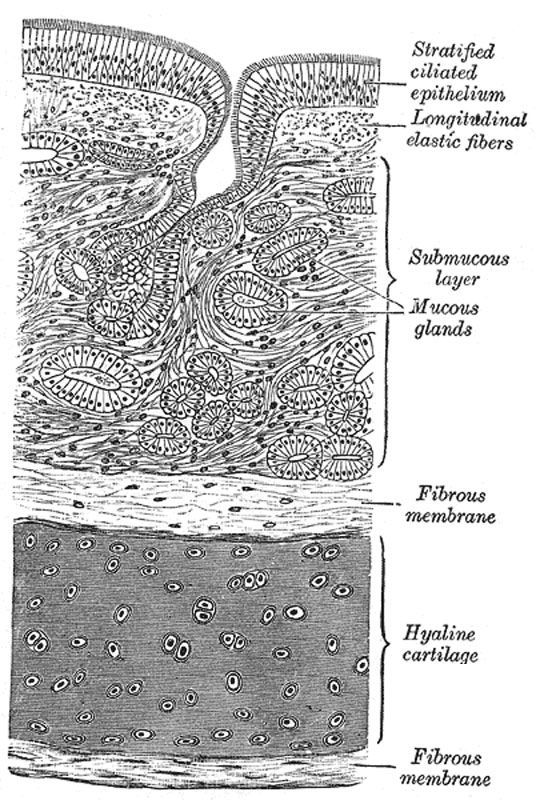
(Fig. 964) The trachea and extrapulmonary bronchi are composed of imperfect rings of hyaline cartilage, fibrous tissue, muscular fibers, mucous membrane, and glands.
The cartilages of the trachea vary from sixteen to twenty in number: each forms an imperfect ring, which occupies the anterior two-thirds or so of the circumference of the trachea, being deficient behind, where the tube is completed by fibrous tissue and unstriped muscular fibers. The cartilages are placed horizontally above each other, separated by narrow intervals. They measure about 4 mm. in depth and 1 mm. in thickness. Their outer surfaces are flattened in a vertical direction, but the internal are convex, the cartilages being thicker in the middle than at the margins. Two or more of the cartilages often unite, partially or completely, and they are sometimes bifurcated at their extremities. They are highly elastic, but may become calcified in advanced life. In the right bronchus the cartilages vary in number from six to eight; in the left, from nine to twelve. They are shorter and narrower than those of the trachea, but have the same shape and arrangement. The peculiar tracheal cartilages are the first and the last (Fig. 961).
The first cartilage is broader than the rest, and often divided at one end; it is connected by the cricotracheal ligament with the lower border of the cricoid cartilage, with which, or with the succeeding cartilage, it is sometimes blended.
The last cartilage is thick and broad in the middle, in consequence of its lower border being prolonged into a triangular hook-shaped process, which curves downward and backward between the two bronchi. It ends on each side in an imperfect ring, which encloses the commencement of the bronchus. The cartilage above the last is somewhat broader than the others at its center.
The Fibrous Membrane
The cartilages are enclosed in an elastic fibrous membrane, which consists of two layers; one, the thicker, passing over the outer surface of the ring, the other over the inner surface: at the upper and lower margins of the cartilages the two layers blend together to form a single membrane, which connects the rings one with another. They are thus invested by the membrane. In the space behind, between the ends of the rings, the membrane forms a single layer.
The muscular tissue consists of two layers of non-striated muscle, longitudinal and transverse. The longitudinal fibers are external, and consist of a few scattered bundles. The transverse fibers (Trachealis muscle) are internal, and form a thin layer which extends transversely between the ends of the cartilages.
Mucous Membrane
The mucous membrane is continuous above with that of the larynx, and below with that of the bronchi. It consists of areolar and lymphoid tissue, and presents a well-marked basement membrane, supporting a stratified epithelium, the surface layer of which is columnar and ciliated, while the deeper layers are composed of oval or rounded cells. Beneath the basement membrane there is a distinct layer of longitudinal elastic fibers with a small amount of intervening areolar tissue. The submucous layer is composed of a loose mesh-work of connective tissue, containing large bloodvessels, nerves, and mucous glands; the ducts of the latter pierce the overlying layers and open on the surface (Fig. 964).
- Gray's Images: Development | Lymphatic | Neural | Vision | Hearing | Somatosensory | Integumentary | Respiratory | Gastrointestinal | Urogenital | Endocrine | Surface Anatomy | iBook | Historic Disclaimer
| Historic Disclaimer - information about historic embryology pages |
|---|
 |
| iBook - Gray's Embryology | |
|---|---|

|
|
Reference
Gray H. Anatomy of the human body. (1918) Philadelphia: Lea & Febiger.
Cite this page: Hill, M.A. (2024, April 27) Embryology Gray0964.jpg. Retrieved from https://embryology.med.unsw.edu.au/embryology/index.php/File:Gray0964.jpg
- © Dr Mark Hill 2024, UNSW Embryology ISBN: 978 0 7334 2609 4 - UNSW CRICOS Provider Code No. 00098G
File history
Click on a date/time to view the file as it appeared at that time.
| Date/Time | Thumbnail | Dimensions | User | Comment | |
|---|---|---|---|---|---|
| current | 23:19, 21 August 2012 | | 536 × 800 (139 KB) | Z8600021 (talk | contribs) | ==Trachea Transverse section== ===Structure=== (Fig. 964) The trachea and extrapulmonary bronchi are composed of imperfect rings of hyaline cartilage, fibrous tissue, muscular fibers, mucous membrane, and glands. The cartilages of the trachea vary from |
You cannot overwrite this file.
File usage
The following 4 pages use this file: