File:Gray0960.jpg

Original file (530 × 650 pixels, file size: 99 KB, MIME type: image/jpeg)
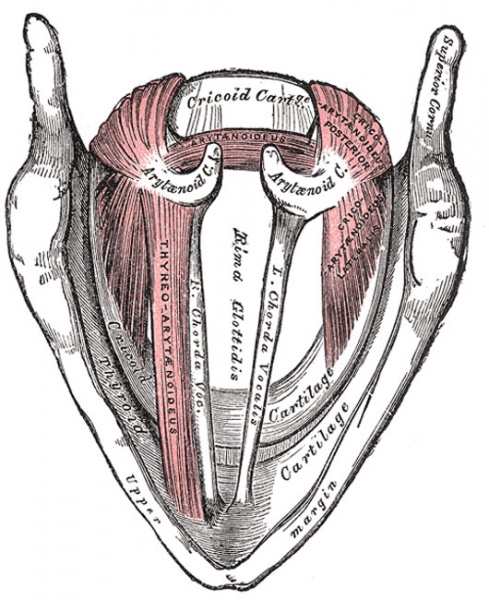
Larynx Muscles
Seen from above.
The Thyreoarytænoideus (Thyroarytenoid) (Figs. 959, 960) is a broad, thin, muscle which lies parallel with and lateral to the vocal fold, and supports the wall of the ventricle and its appendix. It arises in front from the lower half of the angle of the thyroid cartilage, and from the middle cricothyroid ligament. Its fibers pass backward and lateralward, to be inserted into the base and anterior surface of the arytenoid cartilage. The lower and deeper fibers of the muscle can be differentiated as a triangular band which is inserted into the vocal process of the arytenoid cartilage, and into the adjacent portion of its anterior surface; it is termed the Vocalis, and lies parallel with the vocal ligament, to which it is adherent.
A considerable number of the fibers of the Thyreoarytænoideus are prolonged into the aryepiglottic fold, where some of them become lost, while others are continued to the margin of the epiglottis. They have received a distinctive name, Thyreoepiglotticus, and are sometimes described as a separate muscle. A few fibers extend along the wall of the ventricle from the lateral wall of the arytenoid cartilage to the side of the epiglottis and constitute the Ventricularis muscle.
(Text modified from Gray's 1918 Anatomy)
- Larynx Image Links: All cartilages of the larynx | Epiglottis cartilage | Thyroid cartilage | Cricoid cartilage | Arytenoid cartilage | Larynx ligaments anterior | Larynx ligaments posterior | Larynx sagittal section | Larynx and upper trachea | Larynx entrance | Larynx interior | Larynx muscular attachments | Larynx muscles 1 | Larynx muscles 2 | Larynx muscles 3 | Cartilage Development | Respiratory System Development
- Gray's Images: Development | Lymphatic | Neural | Vision | Hearing | Somatosensory | Integumentary | Respiratory | Gastrointestinal | Urogenital | Endocrine | Surface Anatomy | iBook | Historic Disclaimer
| Historic Disclaimer - information about historic embryology pages |
|---|
 |
| iBook - Gray's Embryology | |
|---|---|

|
|
Reference
Gray H. Anatomy of the human body. (1918) Philadelphia: Lea & Febiger.
Cite this page: Hill, M.A. (2024, April 27) Embryology Gray0960.jpg. Retrieved from https://embryology.med.unsw.edu.au/embryology/index.php/File:Gray0960.jpg
- © Dr Mark Hill 2024, UNSW Embryology ISBN: 978 0 7334 2609 4 - UNSW CRICOS Provider Code No. 00098G
File history
Click on a date/time to view the file as it appeared at that time.
| Date/Time | Thumbnail | Dimensions | User | Comment | |
|---|---|---|---|---|---|
| current | 19:02, 21 August 2012 | | 530 × 650 (99 KB) | Z8600021 (talk | contribs) | ==Larynx Muscles== Seen from above. The Thyreoarytænoideus (Thyroarytenoid) (Figs. 959, 960) is a broad, thin, muscle which lies parallel with and lateral to the vocal fold, and supports the wall of the ventricle and its appendix. It arises in front fr |
You cannot overwrite this file.
File usage
The following 4 pages use this file: