File:Gray0893.jpg
Original file (355 × 700 pixels, file size: 93 KB, MIME type: image/jpeg)
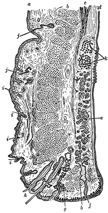
Structure of the Eyelids
Sagittal section through the upper eyelid. (After Waldeyer.)
a. Skin. b. Orbicularis oculi. b’. Marginal fasciculus of Orbicularis (ciliary bundle). c. Levator palpebræ. d. Conjunctiva. e. Tarsus. f. Tarsal gland. g. Sebaceous gland. h. Eyelashes. i. Small hairs of skin. Sweat glands. k. Posterior tarsal glands
=
The eyelids are composed of the following structures taken in their order from without inward: integument, areolar tissue, fibers of the Orbicularis oculi, tarsus, orbital septum, tarsal glands and conjunctiva. The upper eyelid has, in addition, the aponeurosis of the Levator palpebræ superioris (Fig. 893).
The integument is extremely thin, and continuous at the margins of the eyelids with the conjunctiva.
The subcutaneous areolar tissue is very lax and delicate, and seldom contains any fat.
The palpebral fibers of the Orbicularis oculi are thin, pale in color, and possess an involuntary action.
(Text modified from Gray's 1918 Anatomy)
- Gray's Images: Development | Lymphatic | Neural | Vision | Hearing | Somatosensory | Integumentary | Respiratory | Gastrointestinal | Urogenital | Endocrine | Surface Anatomy | iBook | Historic Disclaimer
| Historic Disclaimer - information about historic embryology pages |
|---|
 |
| iBook - Gray's Embryology | |
|---|---|

|
|
Reference
Gray H. Anatomy of the human body. (1918) Philadelphia: Lea & Febiger.
Cite this page: Hill, M.A. (2024, May 21) Embryology Gray0893.jpg. Retrieved from https://embryology.med.unsw.edu.au/embryology/index.php/File:Gray0893.jpg
- © Dr Mark Hill 2024, UNSW Embryology ISBN: 978 0 7334 2609 4 - UNSW CRICOS Provider Code No. 00098G
File history
Click on a date/time to view the file as it appeared at that time.
| Date/Time | Thumbnail | Dimensions | User | Comment | |
|---|---|---|---|---|---|
| current | 22:55, 19 August 2012 | | 355 × 700 (93 KB) | Z8600021 (talk | contribs) | ==Structure of the Eyelids== Sagittal section through the upper eyelid. (After Waldeyer.) a. Skin. b. Orbicularis oculi. b’. Marginal fasciculus of Orbicularis (ciliary bundle). c. Levator palpebræ. d. Conjunctiva. e. Tarsus. f. Tarsal gland. g. Seb |
You cannot overwrite this file.
File usage
The following 2 pages use this file: