File:Gray0883.jpg
Original file (616 × 700 pixels, file size: 107 KB, MIME type: image/jpeg)
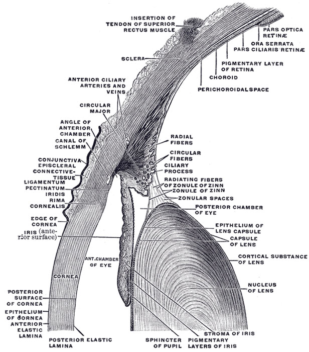
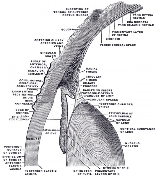
Fig. 883. Section through front of Eyeball
The upper half of a sagittal section through the front of the eyeball.
The Aqueous Humor (humor aqueus) — The aqueous humor fills the anterior and posterior chambers of the eyeball. It is small in quantity, has an alkaline reaction, and consists mainly of water, less than one-fiftieth of its weight being solid matter, chiefly chloride of sodium.
The Vitreous Body (corpus vitreum) — The vitreous body forms about four-fifths of the bulb of the eye. It fills the concavity of the retina, and is hollowed in front, forming a deep concavity, the hyaloid fossa, for the reception of the lens. It is transparent, of the consistence of thin jelly, and is composed of an albuminous fluid enclosed in a delicate transparent membrane, the hyaloid membrane.
The Crystalline Lens (lens crystallina) — The crystalline lens, enclosed in its capsule, is situated immediately behind the iris, in front of the vitreous body, and encircled by the ciliary processes, which slightly overlap its margin.
The Ciliary Body (corpus ciliare) — The ciliary body comprises the orbiculus ciliaris, the ciliary processes, and the Ciliaris muscle.
The Iris — The iris has received its name from its various colors in different individuals. It is a thin, circular, contractile disk, suspended in the aqueous humor between the cornea and lens, and perforated a little to the nasal side of its center by a circular aperture, the pupil.
(Text modified from Gray's 1918 Anatomy)
- Gray's Images: Development | Lymphatic | Neural | Vision | Hearing | Somatosensory | Integumentary | Respiratory | Gastrointestinal | Urogenital | Endocrine | Surface Anatomy | iBook | Historic Disclaimer
| Historic Disclaimer - information about historic embryology pages |
|---|
 |
| iBook - Gray's Embryology | |
|---|---|

|
|
Reference
Gray H. Anatomy of the human body. (1918) Philadelphia: Lea & Febiger.
Cite this page: Hill, M.A. (2024, May 23) Embryology Gray0883.jpg. Retrieved from https://embryology.med.unsw.edu.au/embryology/index.php/File:Gray0883.jpg
- © Dr Mark Hill 2024, UNSW Embryology ISBN: 978 0 7334 2609 4 - UNSW CRICOS Provider Code No. 00098G
File history
Click on a date/time to view the file as it appeared at that time.
| Date/Time | Thumbnail | Dimensions | User | Comment | |
|---|---|---|---|---|---|
| current | 09:33, 19 August 2012 | | 616 × 700 (107 KB) | Z8600021 (talk | contribs) | (Text modified from Gray's 1918 Anatomy) {{Gray Anatomy}} Category:Cartoon Category:Senses Category:Vision Category:Human |
You cannot overwrite this file.
File usage
The following 2 pages use this file: