File:Gray0597.jpg
Gray0597.jpg (700 × 496 pixels, file size: 80 KB, MIME type: image/jpeg)
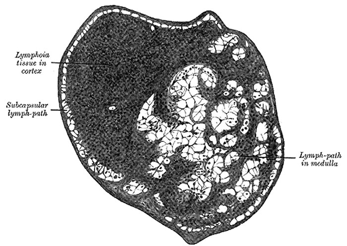
Lymph Gland (Node)
Section of small lymph gland of rabbit. X 100.
| Historic Disclaimer - information about historic embryology pages |
|---|
 |
Structure of Lymph Glands
A lymph gland consists of:
- a fibrous envelope, or capsule, from which a frame-work of processes (trabeculæ) proceeds inward, imperfectly dividing the gland into open spaces freely communicating with each other
- a quantity of lymphoid tissue occupying these spaces without completely filling them
- a free supply of bloodvessels, which are supported in the trabeculæ
- the afferent and efferent vessels communicating through the lymph paths in the substance of the gland.
The nerves passing into the hilus are few in number and are chiefly distributed to the bloodvessels supplying the gland.
The capsule is composed of connective tissue with some plain muscle fibers, and from its internal surface are given off a number of membranous processes or trabeculæ, consisting, in man, of connective tissue, with a small admixture of plain muscle fibers; but in many of the lower animals composed almost entirely of involuntary muscle. They pass inward, radiating toward the center of the gland, for a certain distance—that is to say, for about one-third or one-fourth of the space between the circumference and the center of the gland. In some animals they are sufficiently well-marked to divide the peripheral or cortical portion of the gland into a number of compartments (so-called follicles), but in man this arrangement is not obvious. The larger trabeculæ springing from the capsule break up into finer bands, and these interlace to form a mesh-work in the central or medullary portion of the gland. In these spaces formed by the interlacing trabeculæ is contained the proper gland substance or lymphoid tissue. The gland pulp does not, however, completely fill the spaces, but leaves, between its outer margin and the enclosing trabeculæ, a channel or space of uniform width throughout. This is termed the lymph path or lymph sinus (Fig. 597). Running across it are a number of finer trabeculæ of retiform connective tissue, the fibers of which are, for the most part, covered by ramifying cells.
On account of the peculiar arrangement of the frame-work of the organ, the gland pulp in the cortical portion is disposed in the form of nodules, and in the medullary part in the form of rounded cords. It consists of ordinary lymphoid tissue (Fig. 598), being made up of a delicate net-work of retiform tissue, which is continuous with that in the lymph paths, but marked off from it by a closer reticulation; it is probable, moreover, that the reticular tissue of the gland pulp and the lymph paths is continuous with that of the trabeculæ, and ultimately with that of the capsule of the gland. In its meshes, in the nodules and cords of lymphoid tissue, are closely packed lymph corpuscles. The gland pulp is traversed by a dense plexus of capillary bloodvessels. The nodules or follicles in the cortical portion of the gland frequently show, in their centers, areas where karyokinetic figures indicate a division of the lymph corpuscles. These areas are termed germ centers. The cells composing them have more abundant protoplasm than the peripheral cells.
The afferent vessels, as stated above, enter at all parts of the periphery of the gland, and after branching and forming a dense plexus in the substance of the capsule, open into the lymph sinuses of the cortical part. In doing this they lose all their coats except their endothelial lining, which is continuous with a layer of similar cells lining the lymph paths. In like manner the efferent vessel commences from the lymph sinuses of the medullary portion. The stream of lymph carried to the gland by the afferent vessels thus passes through the plexus in the capsule to the lymph paths of the cortical portion, where it is exposed to the action of the gland pulp; flowing through these it enters the paths or sinuses of the medullary portion, and finally emerges from the hilus by means of the efferent vessel. The stream of lymph in its passage through the lymph sinuses is much retarded by the presence of the reticulum, hence morphological elements, either normal or morbid, are easily arrested and deposited in the sinuses. Many lymph corpuscles pass with the efferent lymph stream to join the general blood stream. The arteries of the gland enter at the hilus, and either go at once to the gland pulp, to break up into a capillary plexus, or else run along the trabeculæ, partly to supply them and partly running across the lymph paths, to assist in forming the capillary plexus of the gland pulp. This plexus traverses the lymphoid tissue, but does not enter into the lymph sinuses. From it the veins commence and emerge from the organ at the same place as that at which the arteries enter.
(Text from Gray's Anatomy 1918)
Gray's Lymphatic Anatomy: 592 Primary lymph sacs | 593 Lymph capillaries of the human conjunctiva | 594 Lymph capillaries from the human scrotum | 595 Lymph capillaries of the sole of the human foot | 596 Section through human tongue | 597 Lymph gland (Node) | 598 Lymph gland tissue | 599 Thoracic and right lymphatic ducts | 600 Modes of origin of thoracic duct | 601 Terminal collecting trunks of right side | 602 Lymph glands of the head | 603 Lymphatics of pharynx | 604 Lymphatics of the face | 605 Lymphatics of the Tongue | 606 Lymph glands of the upper extremity | 607 Lymphatics of the mamma | 608 Lymphatic vessels of the dorsal hand surface | 609 Lymph glands of popliteal fossa | 610 Superficial lymph glands and vessels of the lower extremity | 611 Parietal lymph glands of the pelvis | 612 Iliopelvic glands | 613 Lymphatics of stomach | 614 Lymphatics of stomach | 615 Lymphatics of cecum and vermiform process | 616 Lymphatics of cecum and vermiform process | 617 Lymphatics of Colon | 618 Lymphatic of the Bladder | 619 Lymphatics of the Prostate | 620 Lymphatics of the Uterus | 621 Lymphatics of the thorax and abdomen | 622 Tracheobronchial Lymph Glands | Gray's Anatomy | Historic Disclaimer | Lymphatic Development
- Gray's Images: Development | Lymphatic | Neural | Vision | Hearing | Somatosensory | Integumentary | Respiratory | Gastrointestinal | Urogenital | Endocrine | Surface Anatomy | iBook | Historic Disclaimer
| Historic Disclaimer - information about historic embryology pages |
|---|
|
| iBook - Gray's Embryology | |
|---|---|

|
|
Reference
Gray H. Anatomy of the human body. (1918) Philadelphia: Lea & Febiger.
Cite this page: Hill, M.A. (2024, April 27) Embryology Gray0597.jpg. Retrieved from https://embryology.med.unsw.edu.au/embryology/index.php/File:Gray0597.jpg
- © Dr Mark Hill 2024, UNSW Embryology ISBN: 978 0 7334 2609 4 - UNSW CRICOS Provider Code No. 00098G
File history
Click on a date/time to view the file as it appeared at that time.
| Date/Time | Thumbnail | Dimensions | User | Comment | |
|---|---|---|---|---|---|
| current | 15:15, 14 February 2013 | | 700 × 496 (80 KB) | Z8600021 (talk | contribs) | ==Lymph Node== Section of small lymph gland of rabbit. X 100. {{Gray Anatomy}} Category:Immune Category:Rabbit |
You cannot overwrite this file.
File usage
The following 3 pages use this file: