File:Gray0498.jpg
Gray0498.jpg (475 × 416 pixels, file size: 39 KB, MIME type: image/jpeg)
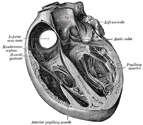
Fig. 498. Section of the Heart showing the Ventricular Septum
The heart consists of muscular fibers, and of fibrous rings which serve for their attachment. It is covered by the visceral layer of the serous pericardium (epicardium), and lined by the endocardium. Between these two membranes is the muscular wall or myocardium.
Endocardium is a thin, smooth membrane which lines and gives the glistening appearance to the inner surface of the heart; it assists in forming the valves by its reduplications, and is continuous with the lining membrane of the large bloodvessels. It consists of connective tissue and elastic fibers, and is attached to the muscular structure by loose elastic tissue which contains bloodvessels and nerves; its free surface is covered by endothelial cells.
Cardiac Muscular Tissue The fibers of the heart differ very remarkably from those of other striped muscles. They are smaller by one-third, and their transverse striæ are by no means so well-marked. They show faint longitudinal striation. The fibers are made up of distinct quadrangular cells, joined end to end so as to form a syncytium (Fig. 499). Each cell contains a clear oval nucleus, situated near its center. The extremities of the cells have a tendency to branch or divide, the subdivisions uniting with offsets from other cells, and thus producing an anastomosis of the fibers. The connective tissue between the bundles of fibers is much less than in ordinary striped muscle, and no sarcolemma has been proved to exist.
Cite this page: Hill, M.A. (2024, April 26) Embryology Gray0498.jpg. Retrieved from https://embryology.med.unsw.edu.au/embryology/index.php/File:Gray0498.jpg
- © Dr Mark Hill 2024, UNSW Embryology ISBN: 978 0 7334 2609 4 - UNSW CRICOS Provider Code No. 00098G
File history
Click on a date/time to view the file as it appeared at that time.
| Date/Time | Thumbnail | Dimensions | User | Comment | |
|---|---|---|---|---|---|
| current | 22:35, 11 October 2009 | | 475 × 416 (39 KB) | S8600021 (talk | contribs) | Section of the heart showing the ventricular septum. |
You cannot overwrite this file.