File:Gray0039.jpg

Original file (1,797 × 1,305 pixels, file size: 464 KB, MIME type: image/jpeg)
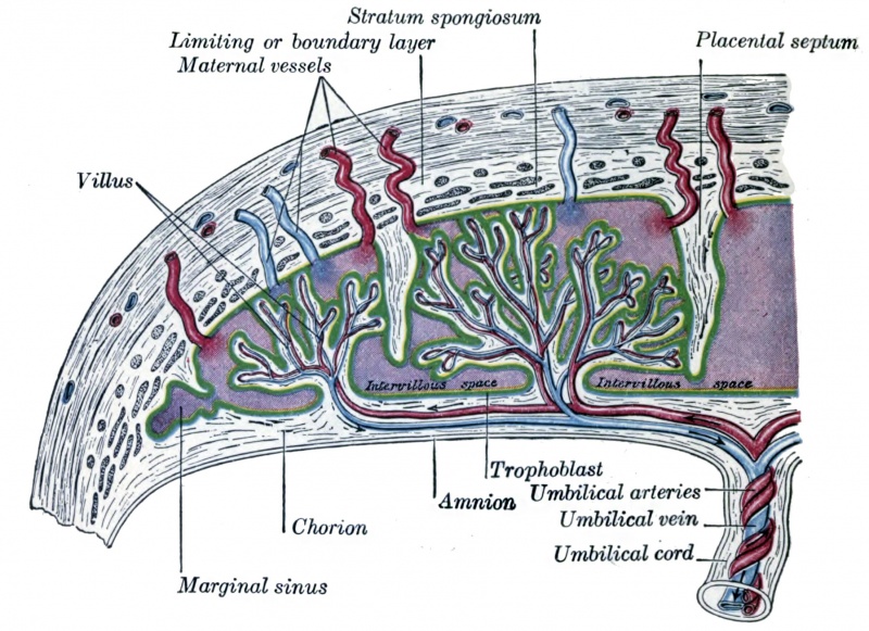
Fig. 39. Scheme of Placental Circulation
- intervillous space - ( choriodecidual space) series of smaller spaces (lacunae) that fuse together to form the maternal blood-filled space.
- trophoblast cells - fetal cells that have invaded the maternal decidua, eroding and opening decidual blood vessels that allows blood to circulate in this space.
- maternal vessels - spiral arteries of the uterine stroma, then decidua, that are modified to remain open into the intervillous space.
- Links: Placenta Development
Fetal Portion
The fetal portion of the placenta consists of the villi of the chorion frondosum; these branch repeatedly, and increase enormously in size. These greatly ramified villi are suspended in the intervillous space, and are bathed in maternal blood, which is conveyed to the space by the uterine arteries and carried away by the uterine veins. A branch of an umbilical artery enters each villus and ends in a capillary plexus from which the blood is drained by a tributary of the umbilical vein. The vessels of the villus are surrounded by a thin layer of mesoderm consisting of gelatinous connective tissue, which is covered by two strata of ectodermal cells derived from the trophoblast: the deeper stratum, next the mesodermic tissue, represents the cytotrophoblast or layer of Langhans; the superficial, in contact with the maternal blood, the syncytiotrophoblast (Figs. 36 and 37). After the fifth month the two strata of cells are replaced by a single layer of somewhat flattened cells.
Maternal Portion
The maternal portion of the placenta is formed by the decidua placentalis containing the intervillous space. As already explained, this space is produced by the enlargement and intercommunication of the spaces in the trophoblastic network. The changes involve the disappearance of the greater portion of the stratum compactum, but the deeper part of this layer persists and is condensed to form what is known as the basal plate. Between this plate and the uterine muscular fibres are the stratum spongiosum and the boundary layer; through these and the basal plate the uterine arteries and veins pass to and from the intervillous space. The endothelial lining of the uterine vessels ceases at the point where they terminate in the intervillous space which is lined by the syncytiotrophoblast. Portions of the stratum compactum persist and are condensed to form a series of septa, which extend from the basal plate through the thickness of the placenta and subdivide it into the lobules or cotyledons seen on the uterine surface of the detached placenta.
- Gray's Images: Development | Lymphatic | Neural | Vision | Hearing | Somatosensory | Integumentary | Respiratory | Gastrointestinal | Urogenital | Endocrine | Surface Anatomy | iBook | Historic Disclaimer
| Historic Disclaimer - information about historic embryology pages |
|---|
 |
| iBook - Gray's Embryology | |
|---|---|

|
|
Reference
Gray H. Anatomy of the human body. (1918) Philadelphia: Lea & Febiger.
Cite this page: Hill, M.A. (2024, April 26) Embryology Gray0039.jpg. Retrieved from https://embryology.med.unsw.edu.au/embryology/index.php/File:Gray0039.jpg
- © Dr Mark Hill 2024, UNSW Embryology ISBN: 978 0 7334 2609 4 - UNSW CRICOS Provider Code No. 00098G
File history
Click on a date/time to view the file as it appeared at that time.
| Date/Time | Thumbnail | Dimensions | User | Comment | |
|---|---|---|---|---|---|
| current | 15:22, 8 June 2014 | | 1,797 × 1,305 (464 KB) | Z8600021 (talk | contribs) | |
| 13:11, 13 August 2012 | 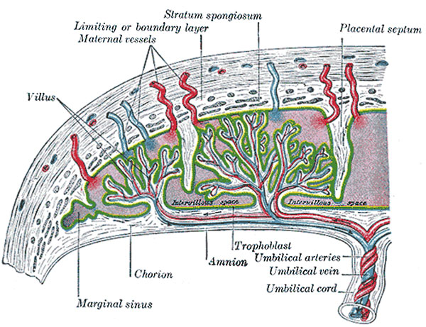 | 600 × 461 (99 KB) | Z8600021 (talk | contribs) | Scheme of placental circulation. Category:Historic Embryology Category:Gray's 1918 Anatomy Category:Human Fetus Category:Placenta |
You cannot overwrite this file.
File usage
The following 10 pages use this file: