File:Gray0027.jpg
From Embryology
No higher resolution available.
Gray0027.jpg (500 × 500 pixels, file size: 39 KB, MIME type: image/jpeg)
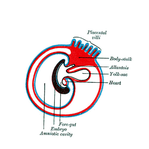
Fig. 27. Diagram showing the expansion of amnion and delimitation of the umbilicus
--Mark Hill (talk) 12:38, 25 April 2013 (EST)
- amnion - An extraembryonic membrane ectoderm and extraembryonic mesoderm in origin and forms the innermost fetal membrane, produces amniotic fluid. This fluid-filled sac initially lies above the trilaminar embryo disc and with embryoic disc folding this sac is drawn ventrally to enclose (cover) the entire embryo, then fetus. The presence of this membane led to the description of reptiles, bird, and mammals as amniotes.
- umbilicus - Term used to describe the navel region, in the embryo anatomically associated with the placental cord, midgut herniation and the allantois.
Early embryo membrane development cartoons: Image 24 | Image 25 | Image 26 | Image 27 | Image 28
Fig 24
Fig 25
Fig 26
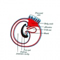
Fig 27
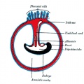
Fig 28


- Gray's Images: Development | Lymphatic | Neural | Vision | Hearing | Somatosensory | Integumentary | Respiratory | Gastrointestinal | Urogenital | Endocrine | Surface Anatomy | iBook | Historic Disclaimer
| Historic Disclaimer - information about historic embryology pages |
|---|
 |
| iBook - Gray's Embryology | |
|---|---|

|
|
Reference
Gray H. Anatomy of the human body. (1918) Philadelphia: Lea & Febiger.
Cite this page: Hill, M.A. (2024, May 4) Embryology Gray0027.jpg. Retrieved from https://embryology.med.unsw.edu.au/embryology/index.php/File:Gray0027.jpg
- © Dr Mark Hill 2024, UNSW Embryology ISBN: 978 0 7334 2609 4 - UNSW CRICOS Provider Code No. 00098G
File history
Click on a date/time to view the file as it appeared at that time.
| Date/Time | Thumbnail | Dimensions | User | Comment | |
|---|---|---|---|---|---|
| current | 11:34, 22 April 2013 | | 500 × 500 (39 KB) | Z8600021 (talk | contribs) |
You cannot overwrite this file.
File usage
The following 15 pages use this file:
- 2009 Lecture 8
- 2010 Lecture 8
- ANAT2341 Lab 4 - Implantation and Villi Development
- ASA Meeting 2013 - Placenta
- Anatomy of the Human Body by Henry Gray
- BGDA Practical Placenta - Implantation and Early Placentation
- Coelomic Cavity Development
- Lecture - Placenta Development
- File:Gray0024-29.gif
- File:Gray0024.jpg
- File:Gray0025.jpg
- File:Gray0026.jpg
- File:Gray0027.jpg
- File:Gray0028.jpg
- Template:Gray fetal membrane cartoons