File:Gray0025.jpg
Gray0025.jpg (500 × 500 pixels, file size: 29 KB, MIME type: image/jpeg)
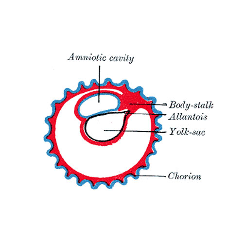
Fig. 25. Diagram illustrating early formation of allantois and differentiation of body-stalk
The Allantois (Figs. 25 to 28).—The allantois arises as a tubular diverticulum of the posterior part of the yolk-sac; when the hind-gut is developed the allantois is carried backward with it and then opens into the cloaca or terminal part of the hind-gut: it grows out into the body-stalk, a mass of mesoderm which lies below and around the tail end of the embryo. The diverticulum is lined by entoderm and covered by mesoderm, and in the latter are carried the allantoic or umbilical vessels. In reptiles, birds, and many mammals the allantois becomes expanded into a vesicle which projects into the extra-embryonic coelom.
--Mark Hill (talk) 12:38, 25 April 2013 (EST)
- allantois - An extraembryonic membrane, endoderm in origin extension from the early hindgut, then cloaca into the connecting stalk of placental animals, connected to the superior end of developing bladder. In reptiles and birds, acts as a reservoir for wastes and mediates gas exchange. In mammals is associated/incorporated with connecting stalk/placental cord fetal-maternal interface.
- body-stalk - (connecting stalk) Historic term used to describe the early extra-embryonic mesoderm region connecting the embryonic disc to the chorion. Present mainly in week 3 of human development, this region will later form the placental cord region, becomes vascular, and connect the embryo to the placenta.
Early embryo membrane development cartoons: Image 24 | Image 25 | Image 26 | Image 27 | Image 28
Fig 24
Fig 25
Fig 26
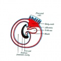
Fig 27
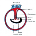
Fig 28


- Gray's Images: Development | Lymphatic | Neural | Vision | Hearing | Somatosensory | Integumentary | Respiratory | Gastrointestinal | Urogenital | Endocrine | Surface Anatomy | iBook | Historic Disclaimer
| Historic Disclaimer - information about historic embryology pages |
|---|
 |
| iBook - Gray's Embryology | |
|---|---|

|
|
Reference
Gray H. Anatomy of the human body. (1918) Philadelphia: Lea & Febiger.
Cite this page: Hill, M.A. (2024, April 26) Embryology Gray0025.jpg. Retrieved from https://embryology.med.unsw.edu.au/embryology/index.php/File:Gray0025.jpg
- © Dr Mark Hill 2024, UNSW Embryology ISBN: 978 0 7334 2609 4 - UNSW CRICOS Provider Code No. 00098G
File history
Click on a date/time to view the file as it appeared at that time.
| Date/Time | Thumbnail | Dimensions | User | Comment | |
|---|---|---|---|---|---|
| current | 11:34, 22 April 2013 | | 500 × 500 (29 KB) | Z8600021 (talk | contribs) |
You cannot overwrite this file.
File usage
The following 15 pages use this file:
- 2009 Lecture 8
- 2010 Lecture 8
- ANAT2341 Lab 4 - Implantation and Villi Development
- ASA Meeting 2013 - Placenta
- Anatomy of the Human Body by Henry Gray
- BGDA Practical Placenta - Implantation and Early Placentation
- Coelomic Cavity Development
- Lecture - Placenta Development
- File:Gray0024-29.gif
- File:Gray0024.jpg
- File:Gray0025.jpg
- File:Gray0026.jpg
- File:Gray0027.jpg
- File:Gray0028.jpg
- Template:Gray fetal membrane cartoons