File:Frazer1910 fig16.jpg

Original file (700 × 745 pixels, file size: 89 KB, MIME type: image/jpeg)
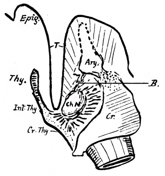
Fig. 16. A semi-diagrammatic sketch from a 22 mm embryo
Showing the chordal nodule, a ready smaller, with its attachments to the cricoid and arytaenoid behind, and central crico-thyroid membrane and lower part of inter-thyroid lamina in front. The front attachments seem stronger than the others, so that these latter thin out and disappear, with the exception of the arytaenoid attachment, which thickens somewhat subsequently and forms the true cord (B).
The arytaenoid ends above in a column of condensed cells, and is joined to the cricoid by similar tissue. T is placed in the transverse cavity and points to the indicating line of -the wall of the cavity.
The inter-thyroid lamina appears to be derived from the ventral lamina in its lowest part: its upper part is presumably formed as a 4th arch structure continuous with the thyroid rudiments.
Online Editor - Embryo CRL 22 mm can be Week 8 Carnegie stage 20 or Carnegie stage 21
| Historic Disclaimer - information about historic embryology pages |
|---|
 |
- Links: fig 1 | fig 2 | fig 3 | fig 4 | fig 5 | fig 6 | fig 7 | fig 8 | fig 9 | fig 10 | fig 11 | fig 12 | fig 13 | fig 14 | fig 15 | fig 16 | fig 17 | fig 18 | fig 19 | 1910 Frazer | Historic Embryology Papers | Respiratory System Development
Reference
Frazer JE. Development of the larynx. (1910) J Anat. 44: 156-191. PMID 17232839
Cite this page: Hill, M.A. (2024, April 27) Embryology Frazer1910 fig16.jpg. Retrieved from https://embryology.med.unsw.edu.au/embryology/index.php/File:Frazer1910_fig16.jpg
- © Dr Mark Hill 2024, UNSW Embryology ISBN: 978 0 7334 2609 4 - UNSW CRICOS Provider Code No. 00098G
File history
Click on a date/time to view the file as it appeared at that time.
| Date/Time | Thumbnail | Dimensions | User | Comment | |
|---|---|---|---|---|---|
| current | 10:17, 12 January 2017 | | 700 × 745 (89 KB) | Z8600021 (talk | contribs) | |
| 17:55, 11 January 2017 | 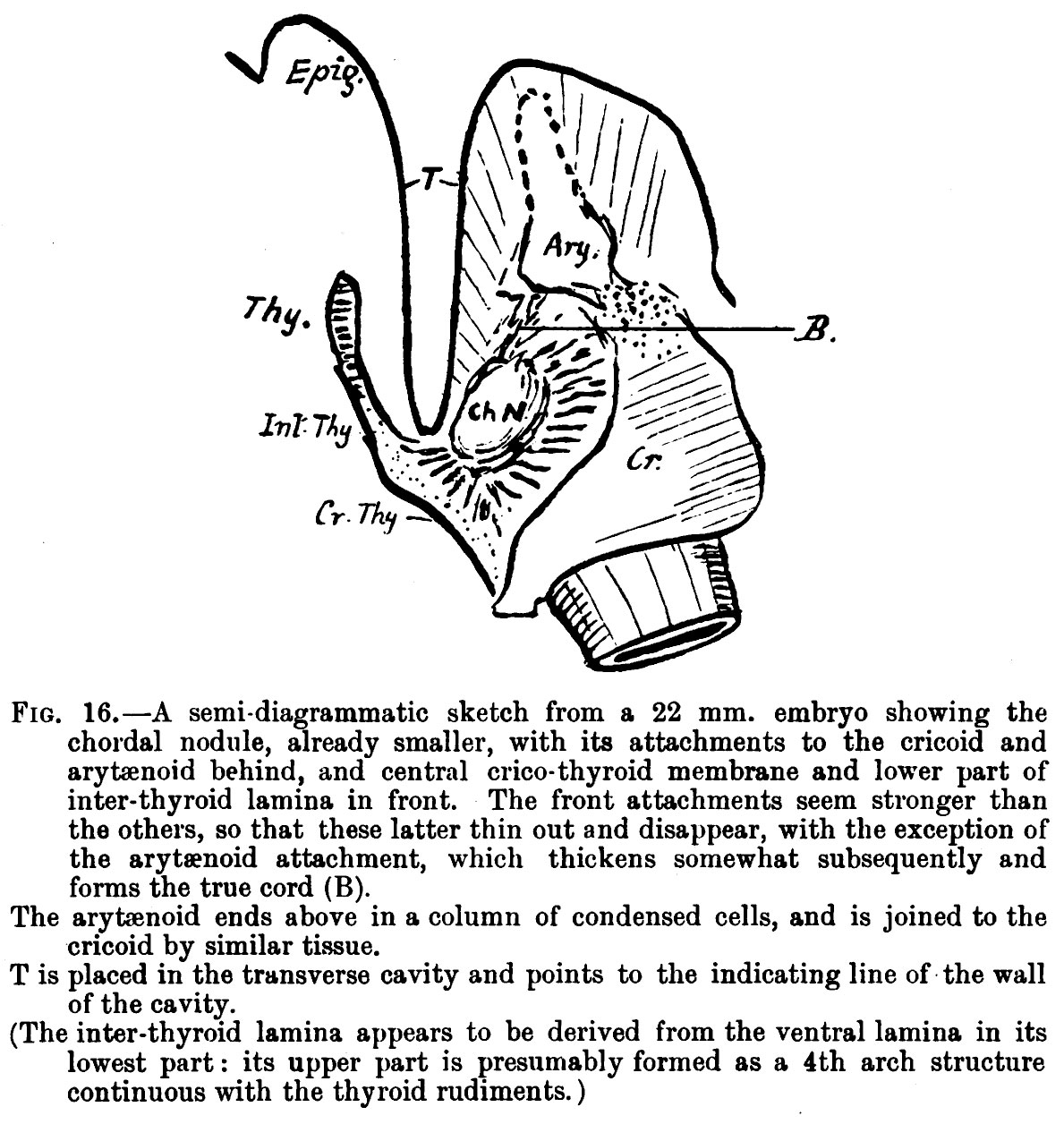 | 1,189 × 1,256 (279 KB) | Z8600021 (talk | contribs) | {{Frazer1910 figures}} |
You cannot overwrite this file.
File usage
The following 2 pages use this file: