File:Frazer1910 fig12.jpg
Original file (1,300 × 450 pixels, file size: 69 KB, MIME type: image/jpeg)
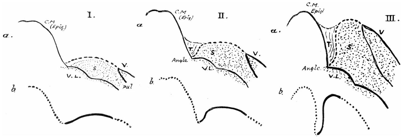
Fig. 12. Diagrams of the mode of formation of the cavity, shown by sagittal sections in the middle line
(a) Diagrams of the mode of formation of the cavity, shown by sagittal sections in the middle line. The central mass (C.M.) is shown in outline, becoming the epiglottis, and the dotted area represents the 5th arch mass: the interrupted line that bounds this above corresponds with the margin of the sagittal opening in the pharyngeal floor. The second and third figures show the result of the overlapping of the central by the lateral mass, thus forming the transverse cavity (T) continuous with the original sagittal cavity (S) round the margin of the latter. The 4th arch element is seen forming the outer part of the dorsal wall of the trans verse cavity. The increase in size of the hinder part (V) of the 5th arch seems to be largely responsible for the lifting forward of the front part. V. L. is the “ ventral lamina” that connects the two 5th arch masses under the floor of the sagittal cavity. The angle of junction between the floors of the two parts of the whole cavity is seen to be well marked.
(b) The lower figures illustrate the change in the direction of the line of the original sagittal opening, which is shown as a. thick line, in the various stages of its alteration.
| Historic Disclaimer - information about historic embryology pages |
|---|
 |
- Links: fig 1 | fig 2 | fig 3 | fig 4 | fig 5 | fig 6 | fig 7 | fig 8 | fig 9 | fig 10 | fig 11 | fig 12 | fig 13 | fig 14 | fig 15 | fig 16 | fig 17 | fig 18 | fig 19 | 1910 Frazer | Historic Embryology Papers | Respiratory System Development
Reference
Frazer JE. Development of the larynx. (1910) J Anat. 44: 156-191. PMID 17232839
Cite this page: Hill, M.A. (2024, April 27) Embryology Frazer1910 fig12.jpg. Retrieved from https://embryology.med.unsw.edu.au/embryology/index.php/File:Frazer1910_fig12.jpg
- © Dr Mark Hill 2024, UNSW Embryology ISBN: 978 0 7334 2609 4 - UNSW CRICOS Provider Code No. 00098G
File history
Click on a date/time to view the file as it appeared at that time.
| Date/Time | Thumbnail | Dimensions | User | Comment | |
|---|---|---|---|---|---|
| current | 09:35, 11 January 2017 | 1,300 × 450 (69 KB) | Z8600021 (talk | contribs) | ||
| 09:34, 11 January 2017 | 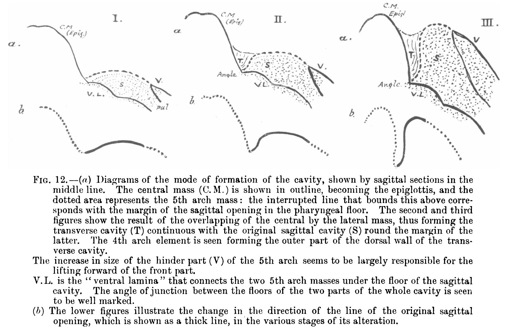 | 1,648 × 1,078 (308 KB) | Z8600021 (talk | contribs) | {{Frazer1910 figures}} |
You cannot overwrite this file.
File usage
The following 2 pages use this file: