File:Frazer1910 fig06.jpg

Original file (1,400 × 1,251 pixels, file size: 319 KB, MIME type: image/jpeg)
Fig. Reconstruction model embryo 6.6 mm
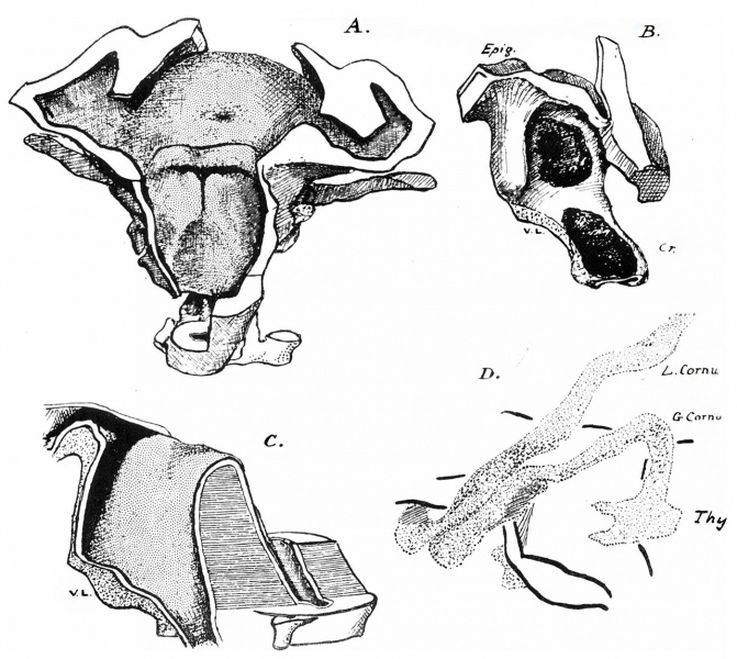
A is a view from behind of the model. about half size, with part of the floor removed below.
B is a side view of the cricoid and arytaanoid rudiments lying against the lining of the segittal cavity: the transverse cavity is seen in front of the arytaenoid. The epiglottic ridge is seen above and in front, and behind this is the lateral mass separated from it by the depression of the transverse opening.
C. Median section through “cavity.” The greater part of the area of the sagittal wall is adherent, leaving only channels along the dorsal wall and floor. The “ ventral lamina. ” (V.L.) is seen below the‘floor, as in the previous figure.
D. The form of the hyoid and thyroid : the lesser cornu is continuous with its fellow in the upper part of the “ body.”
The larynx is outlined deeply. Chondrification seems most advanced in the dorsal aspect of the body, where there is a. single median nodule that is plainly cartilaginous. The rest of the structure shows only precartila.ginous states in various places.
6 A view from behind
6 B side view of the cricoid
6 C Median section through “cavity”
6 D form of the hyoid and thyroid

Online Editor - Embryo CRL 6.6 mm can be Week 5 Carnegie stage 13 or Carnegie stage 14.
| Historic Disclaimer - information about historic embryology pages |
|---|
 |
- Links: fig 1 | fig 2 | fig 3 | fig 4 | fig 5 | fig 6 | fig 7 | fig 8 | fig 9 | fig 10 | fig 11 | fig 12 | fig 13 | fig 14 | fig 15 | fig 16 | fig 17 | fig 18 | fig 19 | 1910 Frazer | Historic Embryology Papers | Respiratory System Development
Reference
Frazer JE. Development of the larynx. (1910) J Anat. 44: 156-191. PMID 17232839
Cite this page: Hill, M.A. (2024, April 28) Embryology Frazer1910 fig06.jpg. Retrieved from https://embryology.med.unsw.edu.au/embryology/index.php/File:Frazer1910_fig06.jpg
- © Dr Mark Hill 2024, UNSW Embryology ISBN: 978 0 7334 2609 4 - UNSW CRICOS Provider Code No. 00098G
File history
Click on a date/time to view the file as it appeared at that time.
| Date/Time | Thumbnail | Dimensions | User | Comment | |
|---|---|---|---|---|---|
| current | 08:59, 11 January 2017 | | 1,400 × 1,251 (319 KB) | Z8600021 (talk | contribs) | |
| 08:57, 11 January 2017 | 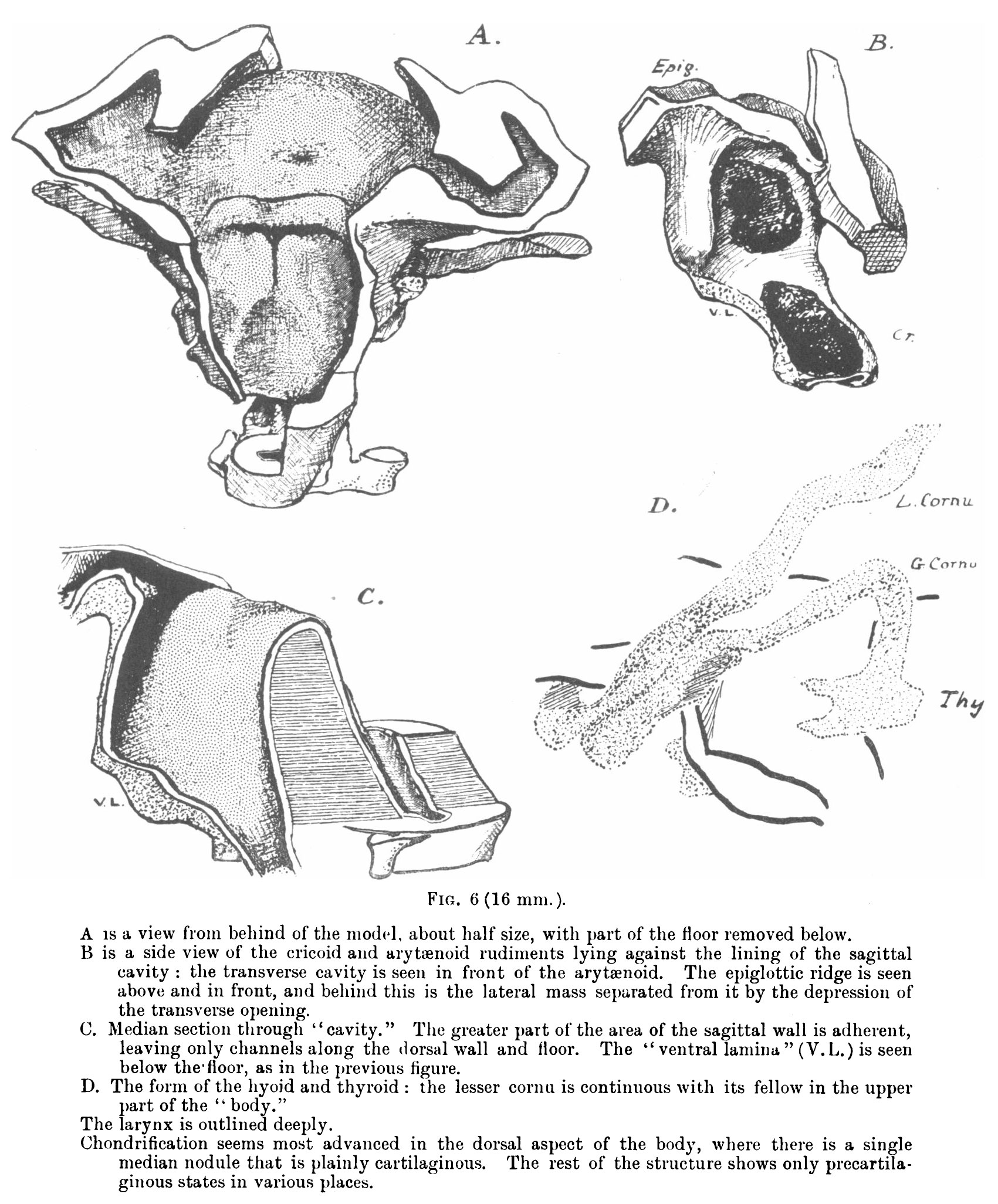 | 1,701 × 2,053 (559 KB) | Z8600021 (talk | contribs) | {{Frazer1910 figures}} |
You cannot overwrite this file.
File usage
The following 4 pages use this file: