File:Foster051.jpg
Original file (722 × 847 pixels, file size: 112 KB, MIME type: image/jpeg)
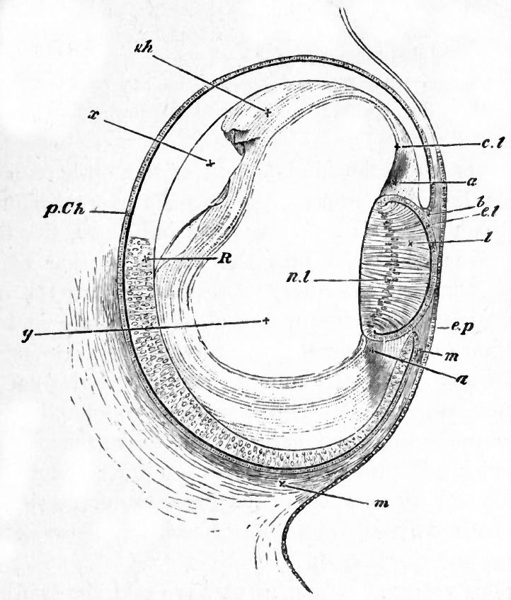
FIG. 51. SECTION OF THE EYE OF CHICK AT THE FOURTH DAY.
ep. superficial epiblast of the side of the head.
R. true retina : anterior wall of the optic cup. p. Gh. pigmentepithelium of the choroid : posterior wall of the optic cup. 6 is placed at the extreme lip of the optic cup at what will become the margin of the iris.
I. the lens. The hind wall, the nuclei of whose elongated cells are shewn at ril, now forms nearly the whole mass of the lens, the front wall being reduced to a layer of flattened cells el.
m. the mesoblast surrounding the optic cup and about to form the choroid and sclerotic. It is seen to pass forward between the lip of the optic cup and the superficial epiblast.
Filling up a large part of the hollow of the optic cup is seen a hyaline mass forming the hyaloid membrane and the coagulum of the vitreous humour. In the neighbourhood of the lens it seems to be continuous as at d with the tissue a, which in turn is continuous with the mesoblast m, and appears to be the rudiment of the capsule of the lens and suspensory ligament.
| Historic Disclaimer - information about historic embryology pages |
|---|
 |
Reference
Foster, M., Balfour, F. M., Sedgwick, A., & Heape, W. (1883). The Elements of Embryology. (2nd ed.). London: Macmillan and Co.
The Elements of Embryology (1883)
File history
Click on a date/time to view the file as it appeared at that time.
| Date/Time | Thumbnail | Dimensions | User | Comment | |
|---|---|---|---|---|---|
| current | 10:24, 11 January 2011 | | 722 × 847 (112 KB) | S8600021 (talk | contribs) | FIG. 51. SECTION OF THE EYE OF CHICK AT THE FOURTH DAY. ep. superficial epiblast of the side of the head. R. true retina : anterior wall of the optic cup. p. Gh. pigmentepithelium of the choroid : posterior wall of the optic cup. 6 is placed at the ex |
You cannot overwrite this file.
File usage
The following page uses this file: