File:Foster047.jpg
Foster047.jpg (484 × 315 pixels, file size: 22 KB, MIME type: image/jpeg)
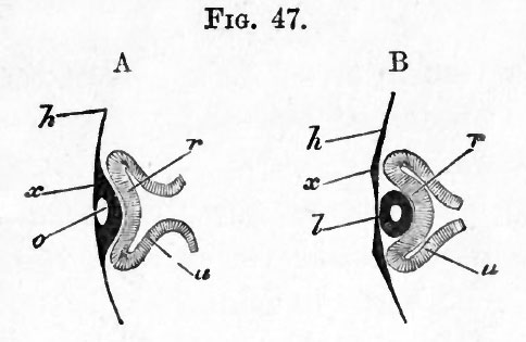
FIG. 47. DIAGRAMMATIC SECTIONS ILLUSTRATING THE FORMATION OF THE EYE. (After Kemak.)
In A, the thin superficial epiblast h is seen to be thickened at #, in front of the optic vesicle, and involuted so as to form a pit o, the mouth of which has already begun to close in. Owing to this involution, which forms the rudiment of the lens, the optic vesicle is doubled in, its front portion r being pushed against the back portion u, and the original cavity of the vesicle thus reduced in size. The stalk of the vesicle is shewn as still broad.
In B, the optic vesicle is still further doubled in so as to form a cup with a posterior wall u and an anterior wall r. In the hollow of this cup lies the lens , now completely detached from the superficial epiblast x. Its cavity is still shewn. The cavity of the stalk of the optic vesicle is already much narrowed. +++++++++++++++++++++++++++
At its first appearance the lens is in immediate contact with the anterior wall of the secondary optic vesicle (Fig. 47 B}. In a short time, however, the lens is seen to lie in the mouth of the cup (Fig. 50 A), a space (vh) (which is occupied by the vitreous humour) making its appearance between the lens and anterior wall of the vesicle.
In order to understand how this space is developed, the position of the optic vesicle and the relations of its stalk must be borne in mind.
The vesicle lies at the side of the head, and its stalk is directed downwards, inwards and backwards. The stalk in fact slants away from the vesicle. Hence when the involution of the lens takes place, the direction in which the front wall of the vesicle is pushed in is not in a line with the axis of the stalk, as for simplicity's sake has been represented in the diagram Fig. 47, but forms an obtuse angle with that axis, after the manner of Fig. 48, where s represents the cavity of the stalk leading away from the almost obliterated cavity of the primary vesicle.
| Historic Disclaimer - information about historic embryology pages |
|---|
 |
Reference
Foster, M., Balfour, F. M., Sedgwick, A., & Heape, W. (1883). The Elements of Embryology. (2nd ed.). London: Macmillan and Co.
The Elements of Embryology (1883)
File history
Click on a date/time to view the file as it appeared at that time.
| Date/Time | Thumbnail | Dimensions | User | Comment | |
|---|---|---|---|---|---|
| current | 10:04, 11 January 2011 | | 484 × 315 (22 KB) | S8600021 (talk | contribs) | FIG. 47. DIAGRAMMATIC SECTIONS ILLUSTRATING THE FORMATION OF THE EYE. (After Kemak.) In A, the thin superficial epiblast h is seen to be thickened at #, in front of the optic vesicle, and involuted so as to form a pit o, the mouth of which has already b |
You cannot overwrite this file.
File usage
The following 2 pages use this file: