File:Foster042.jpg
Foster042.jpg (427 × 378 pixels, file size: 39 KB, MIME type: image/jpeg)
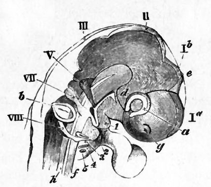
FIG. 42. HEAD OF AN EMBRYO CHICK OF THE THIRD DAY (SEVENTY FIVE HOURS) VIEWED SIDEWAYS AS A TRANSPARENT OBJECT. (From Huxley.)
la. cerebral hemispheres. Ib. vesicle of the third ventricle. II. mid-brain. III. hind-brain, g. nasal pit. a. optic vesicle. 6. otic vesicle, d. infundibulum. e. pineal body. h. notochord. V. fifth nerve. VII. seventh nerve. VIII. united glossopharyngeal and pneumogastric nerves. I, 2, 3, 4, 5 the five visceral folds.
| Historic Disclaimer - information about historic embryology pages |
|---|
 |
Reference
Foster, M., Balfour, F. M., Sedgwick, A., & Heape, W. (1883). The Elements of Embryology. (2nd ed.). London: Macmillan and Co.
The Elements of Embryology (1883)
File history
Click on a date/time to view the file as it appeared at that time.
| Date/Time | Thumbnail | Dimensions | User | Comment | |
|---|---|---|---|---|---|
| current | 09:38, 11 January 2011 | | 427 × 378 (39 KB) | S8600021 (talk | contribs) | FIG. 42. HEAD OF AN EMBRYO CHICK OF THE THIRD DAY (SEVENTY FIVE HOURS) VIEWED SIDEWAYS AS A TRANSPARENT OBJECT. (From Huxley.) la. cerebral hemispheres. Ib. vesicle of the third ventricle. II. mid-brain. III. hind-brain, g. nasal pit. a. optic vesicle. 6 |
You cannot overwrite this file.
File usage
The following 2 pages use this file: