File:Foster024.jpg
From Embryology

Size of this preview: 800 × 362 pixels. Other resolution: 839 × 380 pixels.
Original file (839 × 380 pixels, file size: 54 KB, MIME type: image/jpeg)
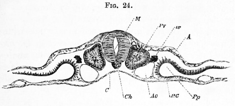
FIG. 24. TRANSVERSE SECTION THROUGH THE DORSAL REGION OP AN EMBRYO OF THE SECOND DAY
(copied from His), introduced here to illustrate the formation of the mesoblastic somite, and the cleavage of the mesoblast.
M. medullary canal ; Pv. mesoblastic somite ; w. rudiment of Wolffian duct; A. epiblast; C. hypoblast ; Ch. notochord ; Ao. aorta ; BO. splanchnopleure.
| Historic Disclaimer - information about historic embryology pages |
|---|
 |
Reference
Foster, M., Balfour, F. M., Sedgwick, A., & Heape, W. (1883). The Elements of Embryology. (2nd ed.). London: Macmillan and Co.
The Elements of Embryology (1883)
File history
Click on a date/time to view the file as it appeared at that time.
| Date/Time | Thumbnail | Dimensions | User | Comment | |
|---|---|---|---|---|---|
| current | 07:27, 9 January 2011 | | 839 × 380 (54 KB) | S8600021 (talk | contribs) | FIG. 24. TRANSVERSE SECTION THROUGH THE DORSAL REGION OP AN EMBRYO OF THE SECOND DAY (copied from His), introduced here to illustrate the formation of the mesoblastic somite, and the cleavage of the mesoblast. M. medullary canal ; Pv. mesoblastic somit |
You cannot overwrite this file.
File usage
The following 2 pages use this file: