File:Fetal rabbit neuroepithelial body 01.jpg
Original file (793 × 1,200 pixels, file size: 98 KB, MIME type: image/jpeg)
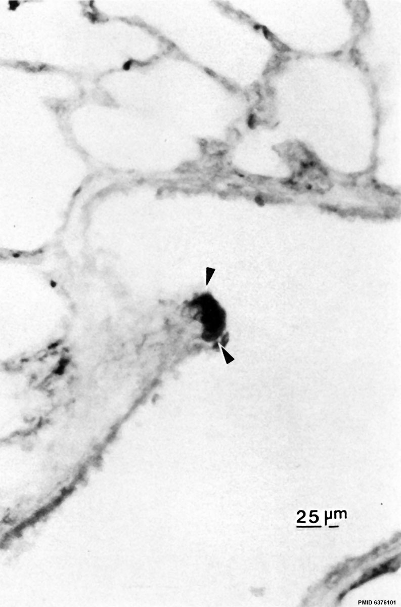
Fetal Rabbit Neuroepithelial Body
Neuroepithelial body (arrows) at an airway bifurcation demonstrated by peroxidase-antiperoxidase (PAP) immunostaining for serotonin, in 29-day fetal rabbit lung.
Paraffin section. Toluidine blue counterstain. x400
Reference
DiAugustine RP & Sonstegard KS. (1984). Neuroendocrinelike (small granule) epithelial cells of the lung. Environ. Health Perspect. , 55, 271-95. PMID: 6376101
Copyright
Copyright: Reproduced with permission from Environmental Health Perspectives (EHP) is a publication of the U.S. Government. Publication of EHP lies in the public domain and is therefore without copyright.
Figure 14. Original image adjusted in size, contrast and labelling.
Cite this page: Hill, M.A. (2024, April 28) Embryology Fetal rabbit neuroepithelial body 01.jpg. Retrieved from https://embryology.med.unsw.edu.au/embryology/index.php/File:Fetal_rabbit_neuroepithelial_body_01.jpg
- © Dr Mark Hill 2024, UNSW Embryology ISBN: 978 0 7334 2609 4 - UNSW CRICOS Provider Code No. 00098G
File history
Click on a date/time to view the file as it appeared at that time.
| Date/Time | Thumbnail | Dimensions | User | Comment | |
|---|---|---|---|---|---|
| current | 10:22, 13 October 2015 | | 793 × 1,200 (98 KB) | Z8600021 (talk | contribs) | |
| 10:21, 13 October 2015 | 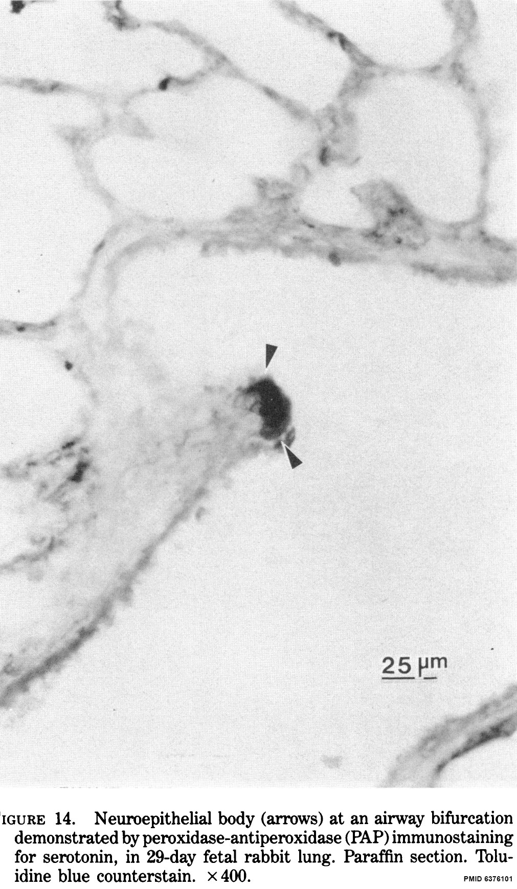 | 1,011 × 1,739 (489 KB) | Z8600021 (talk | contribs) | ==Fetal Rabbit Neuroepithelial Body== Neuroepithelial body (arrows) at an airway bifurcation demonstrated by peroxidase-antiperoxidase (PAP) immunostaining for serotonin, in 29-day fetal rabbit lung. Paraffin section. Toluidine blue counterstain. x40... |
You cannot overwrite this file.
File usage
The following 4 pages use this file: