File:Fetal intra-abdominal umbilical vein varix ultrasound 01.jpg
Fetal_intra-abdominal_umbilical_vein_varix_ultrasound_01.jpg (696 × 242 pixels, file size: 331 KB, MIME type: image/jpeg)
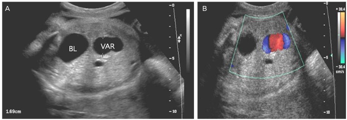
Fetal intra-abdominal umbilical vein varix ultrasound
| (A) Ultrasound transverse view of the lower fetal abdomen | (B) Color Doppler |
|---|---|
| Showing an umbilical vein varix that was approximately 16.9 mm at 33 weeks of gestation. | Shows some turbulence in the intravascular area and differentiates from other cystic lesions. BL, bladder; VAR, fetal umbilical vein varix. |
- Links: placental cord
Reference
Lee SW, Kim MY, Kim JE, Chung JH, Lee HJ & Yoon JY. (2014). Clinical characteristics and outcomes of antenatal fetal intra-abdominal umbilical vein varix detection. Obstet Gynecol Sci , 57, 181-6. PMID: 24883288 DOI.
Copyright
Articles published in Obstet Gynecol Sci are open-access, distributed under the terms of the Creative Commons Attribution Non-Commercial License (http://creativecommons.org/licenses/by-nc/3.0/) which permits unrestricted non-commercial use, distribution, and reproduction in any medium, provided the original work is properly cited.
Ogs-57-181-g001.jpg
File history
Click on a date/time to view the file as it appeared at that time.
| Date/Time | Thumbnail | Dimensions | User | Comment | |
|---|---|---|---|---|---|
| current | 15:49, 1 June 2019 | 696 × 242 (331 KB) | Z8600021 (talk | contribs) | {| | (A) Transverse view of the lower fetal abdomen showing an umbilical vein varix that was approximately 16.9 mm at 33 weeks of gestation. | (B) Color Doppler shows some turbulence in the intravascular area and differentiates from other cystic lesio... |
You cannot overwrite this file.
File usage
The following page uses this file: