File:Fetal integumentary histology Adult.jpg
Fetal_integumentary_histology_Adult.jpg (259 × 217 pixels, file size: 30 KB, MIME type: image/jpeg)
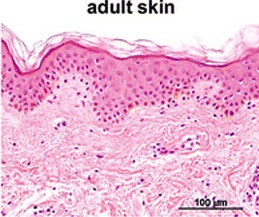
Adult Integumentary Histology- contained basal, spinous, granular and cornified layers.
Scale bars 100 μm
Reference
<pubmed>19701759</pubmed>| PMC2799629 | Arch Dermatol Res
© Coolen NA, Schouten KC, Middelkoop E, Ulrich MM. 2009 Open Access - This article is distributed under the terms of the Creative Commons Attribution Noncommercial License which permits any noncommercial use, distribution, and reproduction in any medium, provided the original author(s) and source are credited.
Original file name: Fig. 1 403_2009_989_Fig1_HTML.gif
--Mark Hill (talk) 11:30, 7 November 2014 (EST) Assessment - Figure relates to project topic contains reference, copyright and student template. Figure is far to small to see any detailed histology, a better image could have been used here.
- Note - This image was originally uploaded as part of an undergraduate science student project and may contain inaccuracies in either description or acknowledgements. Students have been advised in writing concerning the reuse of content and may accidentally have misunderstood the original terms of use. If image reuse on this non-commercial educational site infringes your existing copyright, please contact the site editor for immediate removal.
File history
Click on a date/time to view the file as it appeared at that time.
| Date/Time | Thumbnail | Dimensions | User | Comment | |
|---|---|---|---|---|---|
| current | 23:05, 7 October 2014 | | 259 × 217 (30 KB) | Z3418488 (talk | contribs) |
You cannot overwrite this file.
File usage
The following 2 pages use this file: