File:Fawcett 1910 fig15.jpg
Original file (962 × 973 pixels, file size: 200 KB, MIME type: image/jpeg)
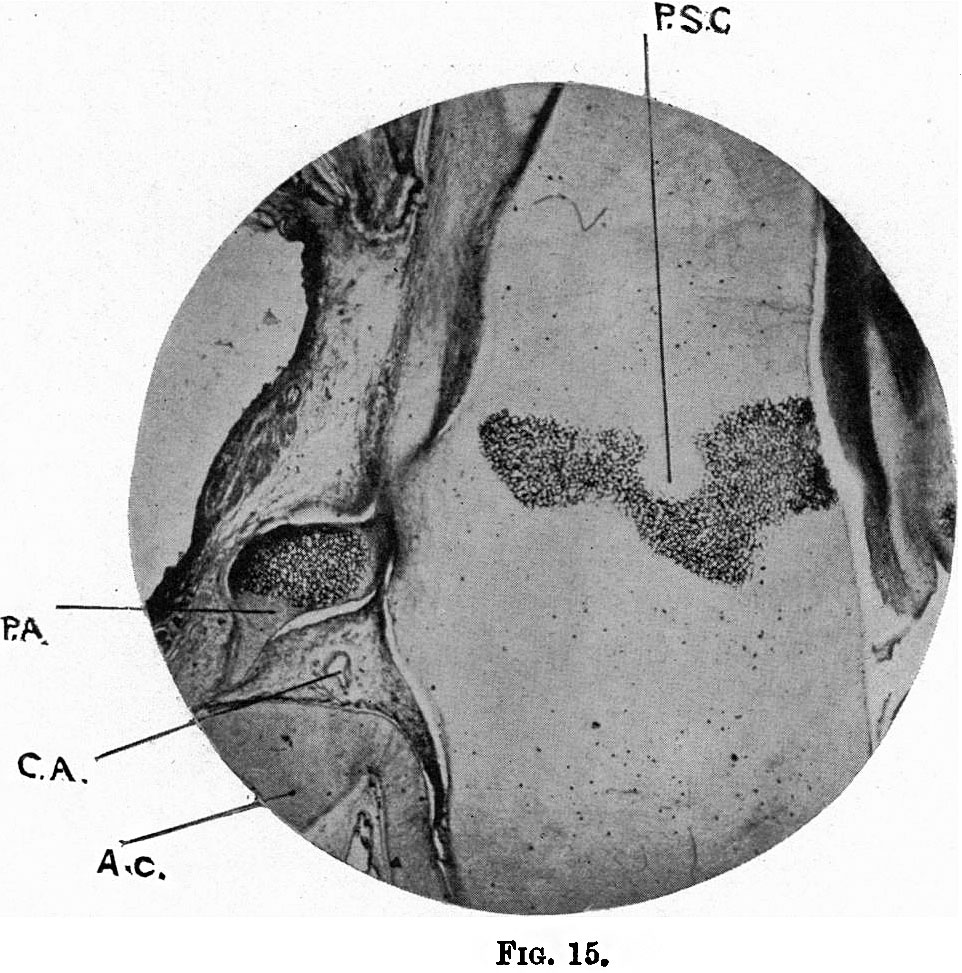
Fig. 15.Photograph of a horizontal section of the head of a 110 mm embryo
Shows two points of great interest, viz. ossification in the corpus sphenoidale - the post-sphenoidal centre (P.S.C.), which has a somewhat curious form,and giving rise to a litle doubt as to whether it is a single centre or the result of fusion of two originally independent ones; 1 and ossification in the processus alaris (P.A.). This is obviously independent of that in the corpus sphenoidale,and its large size is somewhat surprising, if it form only that part commonly in our text-books called lingula. From its posterior end a pointed cartilaginous process is seen to pass backwards towards the auditory capsule (A.C.), and between the processus alaris and the auditory capsule the internal carotid artery (C.A.) is seen in transverse section.
| Historic Disclaimer - information about historic embryology pages |
|---|
 |
|
|
Reference
Fawcett E. Notes on the development of the human sphenoid. (1910) J Anat. Physiol. 44(3): 207-22. PMID 17232842
Cite this page: Hill, M.A. (2024, April 27) Embryology Fawcett 1910 fig15.jpg. Retrieved from https://embryology.med.unsw.edu.au/embryology/index.php/File:Fawcett_1910_fig15.jpg
- © Dr Mark Hill 2024, UNSW Embryology ISBN: 978 0 7334 2609 4 - UNSW CRICOS Provider Code No. 00098G
File history
Click on a date/time to view the file as it appeared at that time.
| Date/Time | Thumbnail | Dimensions | User | Comment | |
|---|---|---|---|---|---|
| current | 08:34, 29 December 2014 | | 962 × 973 (200 KB) | Z8600021 (talk | contribs) |
You cannot overwrite this file.
File usage
The following page uses this file: