File:Fawcett 1910 fig08.jpg
Original file (1,087 × 1,050 pixels, file size: 261 KB, MIME type: image/jpeg)
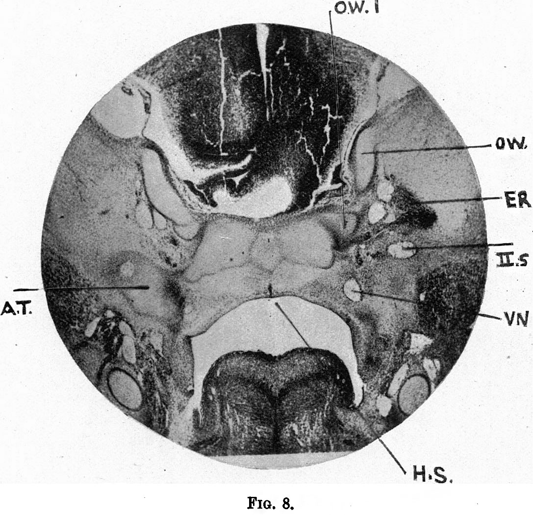
Fig.8. Coronal section of a 19 mm Embryo
Coronal section further forward than fig.7, showing (O.W.1) the posterior limb of the orbital wing which bounds posteriorly the optic foramen. The anterior limb is not yet chondrified. Between O.W. and O.W.1 the outgoing optic stalk is evident; H.S. is the hypophysis stalk; above it is a curious middle piece to the corpus sphenoidale, which, though appearing to be independent, is not so, as models prove. A.T.is the ala temporalis (greater wing); I.5, the 2nd branch of the Trigeminus; E.R.the external rectus. The chief interest in the figure is the independence of the orbital wing (O.W.1).
| Historic Disclaimer - information about historic embryology pages |
|---|
 |
|
|
Reference
Fawcett E. Notes on the development of the human sphenoid. (1910) J Anat. Physiol. 44(3): 207-22. PMID 17232842
Cite this page: Hill, M.A. (2024, April 27) Embryology Fawcett 1910 fig08.jpg. Retrieved from https://embryology.med.unsw.edu.au/embryology/index.php/File:Fawcett_1910_fig08.jpg
- © Dr Mark Hill 2024, UNSW Embryology ISBN: 978 0 7334 2609 4 - UNSW CRICOS Provider Code No. 00098G
File history
Click on a date/time to view the file as it appeared at that time.
| Date/Time | Thumbnail | Dimensions | User | Comment | |
|---|---|---|---|---|---|
| current | 08:33, 29 December 2014 | | 1,087 × 1,050 (261 KB) | Z8600021 (talk | contribs) | {{Fawcett1910_sphenoid_figures}} |
You cannot overwrite this file.
File usage
The following page uses this file: