File:Evans1909a fig01.jpg
Original file (653 × 1,280 pixels, file size: 128 KB, MIME type: image/jpeg)
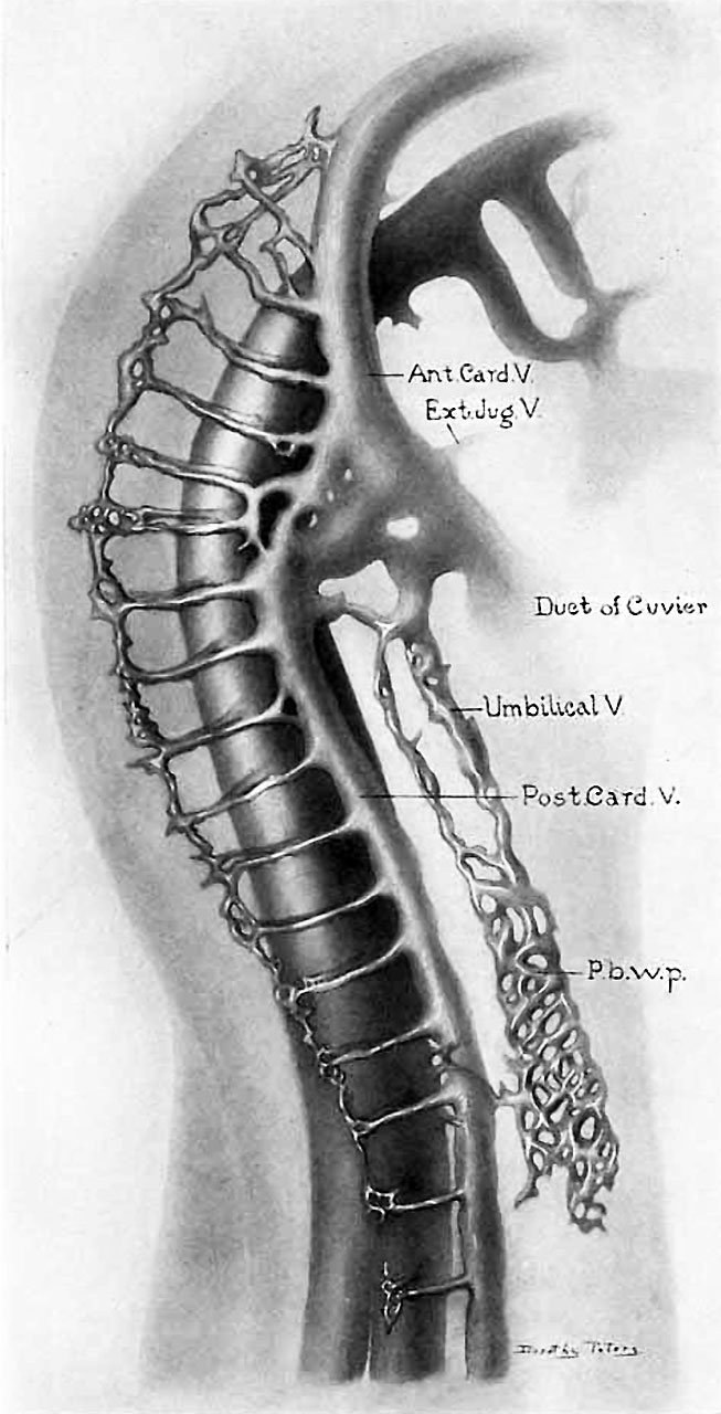
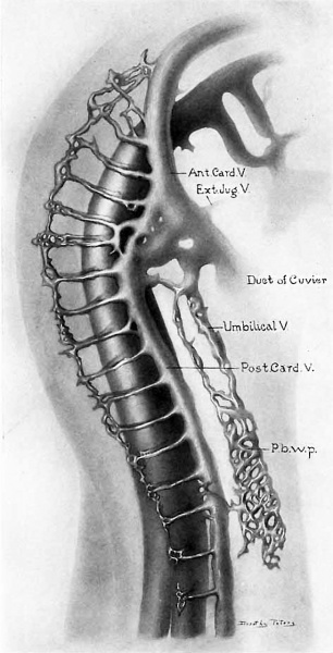
Fig. 1. Chick embryo of 30 somites
(embryo 2 of table). Showing downgrowth of primary wall plexus, x 53.5. Ant. Card. V., anterior cardinal vein. Ext. Jug. V.. external or inferior jugular vein (linguo-facial vein). Post Card. V., posterior cardinal vein. P. b. w. p., primary body wall plexus.
Reference
Evans HM. On the earliest blood-vessels in the anterior limb-buds of birds and their relation to the primary subclavian artery. (1909) Amer. J Anat. 9: 281-319.
Cite this page: Hill, M.A. (2024, April 27) Embryology Evans1909a fig01.jpg. Retrieved from https://embryology.med.unsw.edu.au/embryology/index.php/File:Evans1909a_fig01.jpg
- © Dr Mark Hill 2024, UNSW Embryology ISBN: 978 0 7334 2609 4 - UNSW CRICOS Provider Code No. 00098G
File history
Click on a date/time to view the file as it appeared at that time.
| Date/Time | Thumbnail | Dimensions | User | Comment | |
|---|---|---|---|---|---|
| current | 14:29, 10 August 2017 | | 653 × 1,280 (128 KB) | Z8600021 (talk | contribs) | |
| 14:29, 10 August 2017 | 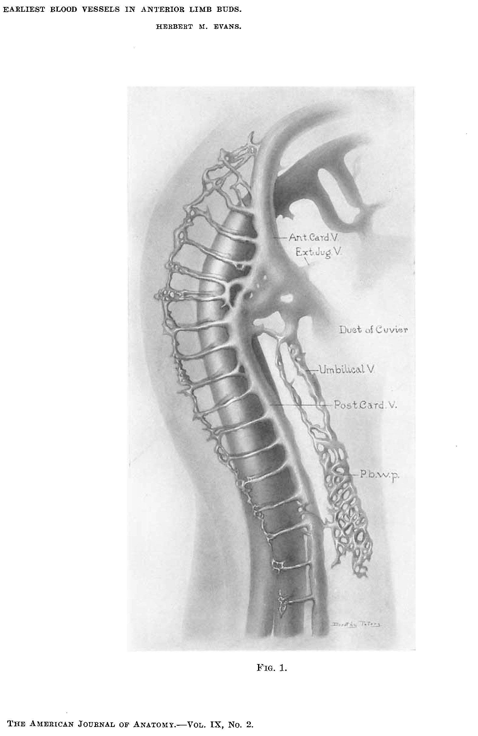 | 1,580 × 2,435 (245 KB) | Z8600021 (talk | contribs) | ===Reference=== {{Ref-Evans1909a}} |
You cannot overwrite this file.
File usage
The following page uses this file: