File:Evans1909 fig10.jpg

Original file (800 × 1,298 pixels, file size: 47 KB, MIME type: image/jpeg)
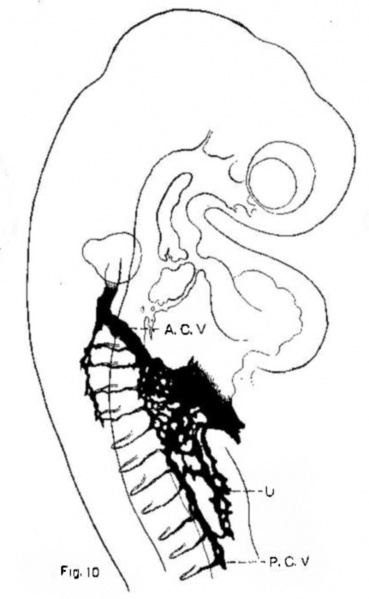
Fig. 10. Injected chick embryo of 24 somites
To show the extension in the somatopleure, of capillary plexus forming the umbilical vein.
A. C. V. = anterior cardinal vein; P. C. V. = posterlor cardinal vein; U = capiliaries destined to form the umbilical vein.
| Historic Disclaimer - information about historic embryology pages |
|---|
 |
Evans (1909) Figures: 1 Chick 17 somites | 2 Chick 20 somites | 3 Chick 23 somites | 6 Chick lateral 25 somites |
Reference
Evans HM. On the development of the aortae, cardinal and umbilical veins, and the other blood vessels of vertebrate embryos from capillaries. (1909) Anat. Rec. 3: 498-518.
Cite this page: Hill, M.A. (2024, May 11) Embryology Evans1909 fig10.jpg. Retrieved from https://embryology.med.unsw.edu.au/embryology/index.php/File:Evans1909_fig10.jpg
- © Dr Mark Hill 2024, UNSW Embryology ISBN: 978 0 7334 2609 4 - UNSW CRICOS Provider Code No. 00098G
File history
Click on a date/time to view the file as it appeared at that time.
| Date/Time | Thumbnail | Dimensions | User | Comment | |
|---|---|---|---|---|---|
| current | 08:03, 29 November 2017 | | 800 × 1,298 (47 KB) | Z8600021 (talk | contribs) |
You cannot overwrite this file.
File usage
There are no pages that use this file.