File:Early Heart Tube (Dorsal).jpg
From Embryology
Size of this preview: 684 × 600 pixels. Other resolution: 1,282 × 1,124 pixels.
Original file (1,282 × 1,124 pixels, file size: 111 KB, MIME type: image/jpeg)
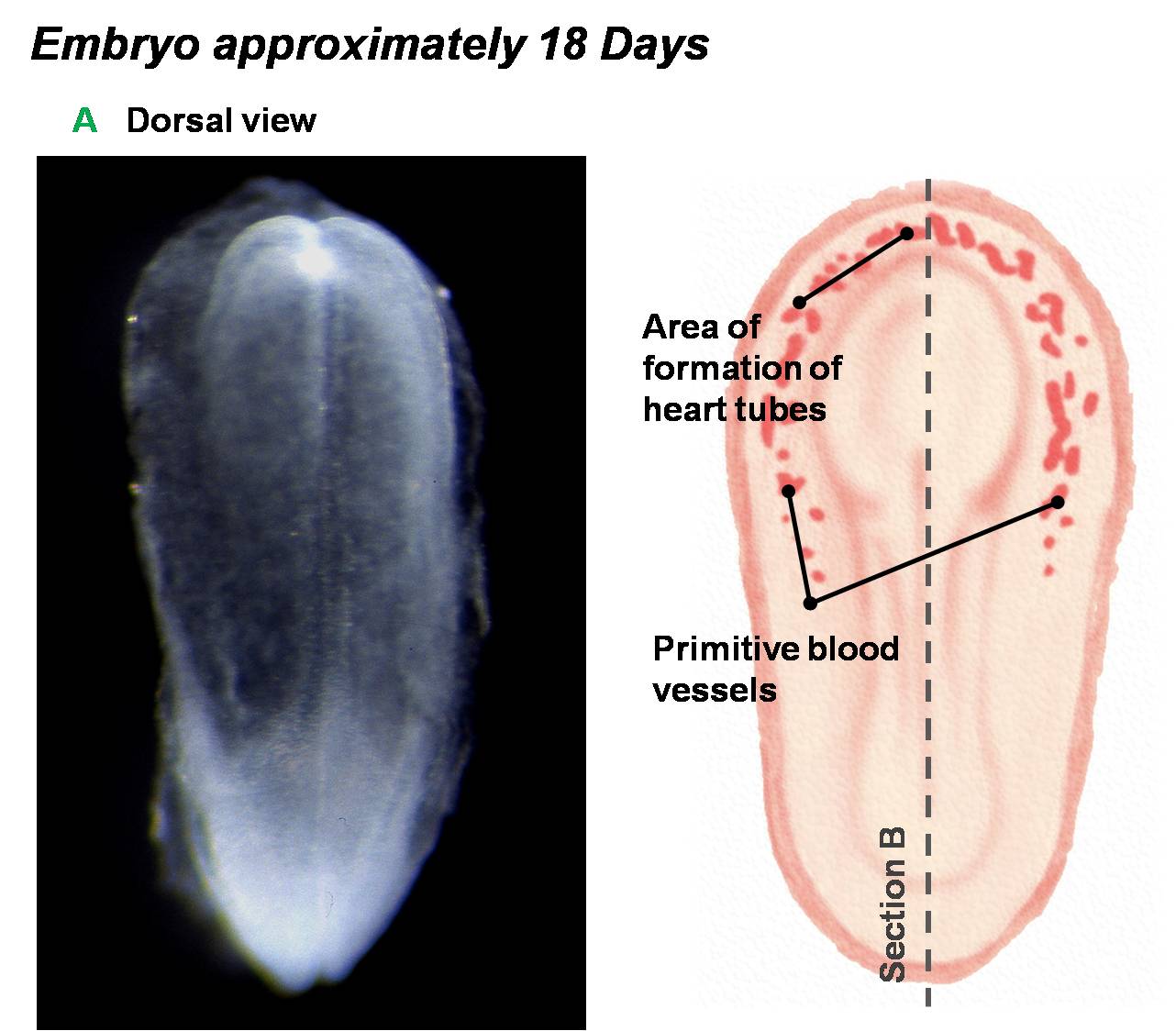
Angiogenesis throughout the embryo allows for the development of angioblastic cords in the cardiogenic mesoderm of the embryo.
Image Source: Scanning electron micrographs of the Carnegie stages of the early human embryos are reproduced with the permission of Prof Kathy Sulik, from embryos collected by Dr. Vekemans and Tania Attié-Bitach. Images are for educational purposes only and cannot be reproduced electronically or in writing without permission.
File history
Click on a date/time to view the file as it appeared at that time.
| Date/Time | Thumbnail | Dimensions | User | Comment | |
|---|---|---|---|---|---|
| current | 10:37, 14 March 2010 | | 1,282 × 1,124 (111 KB) | Z3212774 (talk | contribs) | category:Heart ILP Angiogenesis throughout the embryo allows for the development of angioblastic cords in the cardiogenic mesoderm of the embryo. |
You cannot overwrite this file.
File usage
The following file is a duplicate of this file (more details):
.jpg)
.jpg&oldid=21495)