File:Dickie1914 fig04.jpg
From Embryology
No higher resolution available.
Dickie1914_fig04.jpg (755 × 578 pixels, file size: 71 KB, MIME type: image/jpeg)
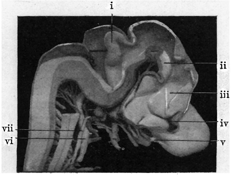
Fig. 4. Shows medial aspect of model of brain
i, Cerebellum
ii, tegmontum
iii, thalamus
iv, Interventriculsr foramen
v, olfactory bulb
vi, vagus
vii, sympathetic
| Historic Disclaimer - information about historic embryology pages |
|---|
 |
Reference
Dickie JK. The anatomy of the head end of a 20 mm human embryo. (1914) J Anat Physiol., 48(4): 445-60. PMID 17233010
Cite this page: Hill, M.A. (2024, April 27) Embryology Dickie1914 fig04.jpg. Retrieved from https://embryology.med.unsw.edu.au/embryology/index.php/File:Dickie1914_fig04.jpg
- © Dr Mark Hill 2024, UNSW Embryology ISBN: 978 0 7334 2609 4 - UNSW CRICOS Provider Code No. 00098G
File history
Click on a date/time to view the file as it appeared at that time.
| Date/Time | Thumbnail | Dimensions | User | Comment | |
|---|---|---|---|---|---|
| current | 21:50, 19 August 2015 | | 755 × 578 (71 KB) | Z8600021 (talk | contribs) | |
| 21:48, 19 August 2015 | 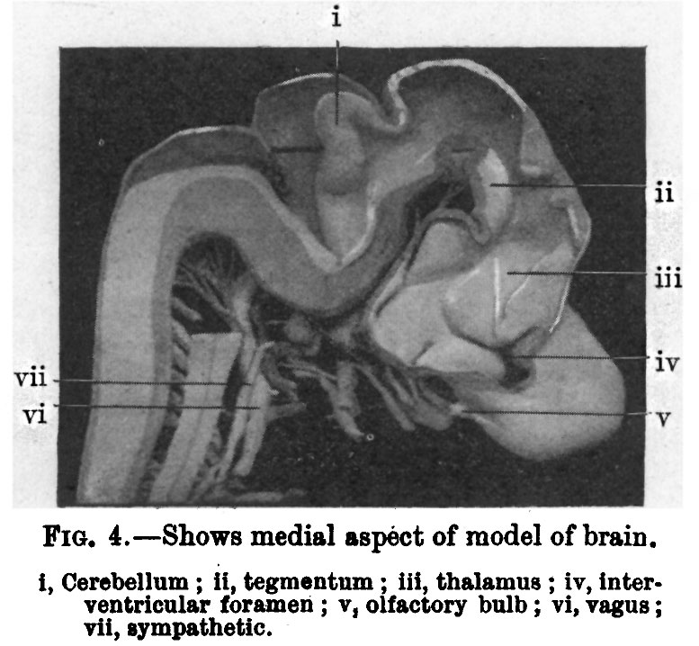 | 777 × 718 (99 KB) | Z8600021 (talk | contribs) | {{Dickie1914 figures}} |
You cannot overwrite this file.
File usage
The following 2 pages use this file: