File:Cullen1916 fig02.jpg
Original file (1,280 × 1,110 pixels, file size: 403 KB, MIME type: image/jpeg)
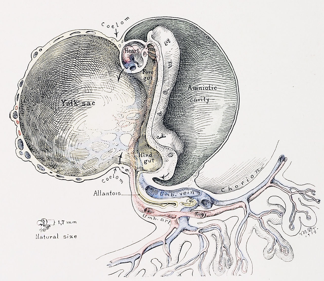
Fig. 2. A More Advanced Stage in the Formation of the Umbilical Region
(Human embryo, 1.7 mm. long. - Mall series, 391.) (This embryo has been very carefully described by Dr. Walter E. Dandy (Amer. Jour. Anat., 1910, x, 85).
Note the advancing approach of the cranial and caudal portions of the yolk-sac and its division into a main cavity and two recesses, the fore-gut and the hind-gut, into the latter of which the allantois now opens. The vitelline arteries and veins are clearly seen on the embryonic side of the yolk-sac. The amnion is now gradually enveloping the embryo. Compare the situation of the coelom in this with that in the subsequent pictures. There is as yet no umbilical cord. For the first stage of its development, See Fig. 3. folding of the exocoe1om. The arrows indicate the direction that it will follow later.
| Historic Disclaimer - information about historic embryology pages |
|---|
 |
- Figure Links: 1 Human embryo 0.7 mm | 2 Human embryo 1.7 mm | 3 Human embryo 2.5 mm | 4 Human embryo 3.5 mm | 5 Human embryo 5 mm | 6 Human embryo 7 mm | 7 Human embryo 7 mm | 8 Human embryo 10 mm | 9 Human embryo 12.5 mm | 10 Human embryo 10 mm | 11 Human embryo 23 mm | 12 Human embryo 3 cm | 13 Human embryo 4.5 cm sagittal | 14 Human Embryo 4.5 cm | 15 Human Embryo 5.2 cm | 16 Human Embryo 6.5 cm | 17 Human Embryo 7.5 cm | 18 Human Embryo 9 cm | 19 Human Embryo 10 cm | 20 Human Embryo 12 cm | 21 Human Embryo 12 cm | 22 Human Embryo 12 cm | 23 Human Embryo 12 cm Cord | 28 Fetus Five Months | 30 Ventral Heria | 31 Human Embryo 5.5 cm | 32 Term Human | 33 Term Human | [[Figures
Reference
Cullen TS. Embryology, anatomy, and diseases of the umbilicus together with diseases of the urachus. (1916) W. B. Saunders Company, Philadelphia And London.
Cite this page: Hill, M.A. (2024, May 13) Embryology Cullen1916 fig02.jpg. Retrieved from https://embryology.med.unsw.edu.au/embryology/index.php/File:Cullen1916_fig02.jpg
- © Dr Mark Hill 2024, UNSW Embryology ISBN: 978 0 7334 2609 4 - UNSW CRICOS Provider Code No. 00098G
File history
Click on a date/time to view the file as it appeared at that time.
| Date/Time | Thumbnail | Dimensions | User | Comment | |
|---|---|---|---|---|---|
| current | 15:58, 27 October 2018 | | 1,280 × 1,110 (403 KB) | Z8600021 (talk | contribs) | |
| 15:57, 27 October 2018 |  | 2,149 × 1,622 (686 KB) | Z8600021 (talk | contribs) | Fig. 2. — A More Advanced Stage in the Formation of the Umbilical Region. (Human embryo, 1.7 mm. long. - Mall series, {{CE391}}.) (This embryo has been very carefully described by Dr. Walter E. Dandy (Amer. Jour. Anat., 1910, x, 85). Note the advan... |
You cannot overwrite this file.
File usage
The following 3 pages use this file: