File:Corner1922 fig01.jpg
From Embryology
No higher resolution available.
Corner1922_fig01.jpg (718 × 597 pixels, file size: 81 KB, MIME type: image/jpeg)
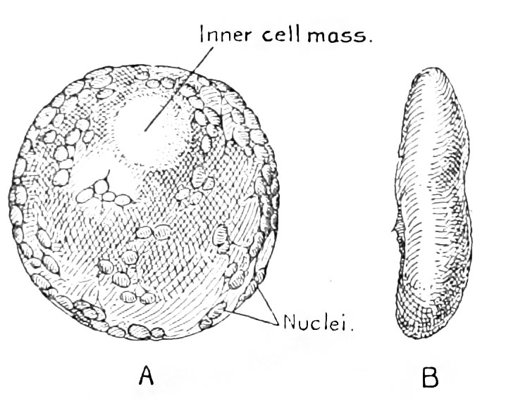
Fig. 1. A Normal and a Pathological Ovum
A normal (A) and a pathological (B) ovum obtained from the same uterus. The more detailed structure of these specimens is shown in figure 7, plate 2. The pathological ovum is wrinkled and compressed. Here it is shown from a side view; in figure 7 its broader surface is shown.
| Historic Disclaimer - information about historic embryology pages |
|---|
 |
File history
Click on a date/time to view the file as it appeared at that time.
| Date/Time | Thumbnail | Dimensions | User | Comment | |
|---|---|---|---|---|---|
| current | 18:25, 13 April 2015 | | 718 × 597 (81 KB) | Z8600021 (talk | contribs) | |
| 18:24, 13 April 2015 |  | 1,000 × 458 (89 KB) | Z8600021 (talk | contribs) | ==Fig. 1. A Normal and a Pathological Ovum== A normal (A) and a pathological (B) ovum obtained from the same uterus. The more detailed structure of these specimens is shown in figure 7, plate 2. The pathological ovum is wrinkled and compressed. Here i... |
You cannot overwrite this file.
File usage
The following page uses this file: