File:Cardiac Muscle EM05.jpg

Original file (992 × 733 pixels, file size: 158 KB, MIME type: image/jpeg)
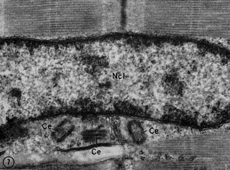
Cardiac Muscle Electron Micrograph
This is a historic (1969) EM showing key features in cardiac muscle ultrastructure. (Stain - Osmium)
A portion of the nucleus of a cardiac muscle cell showing the peripheral dis- tribution of the chromatin typical of glutaraldehyde-fixed muscle . Adjacent to the nucleus is a small portion of the Golgi complex and three centrioles (Ce). Since centrioles generally occur in pairs, it is assumed that a fourth centriole is present but out of the plane of section.
Legend
- Ncl - nucleus.
- Ce - centrioles
Original image X 42,000
Reference
<pubmed>4891913</pubmed>| PMC2107571
Copyright
Rockefeller University Press - Copyright Policy This article is distributed under the terms of an Attribution–Noncommercial–Share Alike–No Mirror Sites license for the first six months after the publication date (see http://www.jcb.org/misc/terms.shtml). After six months it is available under a Creative Commons License (Attribution–Noncommercial–Share Alike 4.0 Unported license, as described at https://creativecommons.org/licenses/by-nc-sa/4.0/ ). (More? Help:Copyright Tutorial)
Original article figure (FIG. 1) has been scaled and rotated.
File history
Click on a date/time to view the file as it appeared at that time.
| Date/Time | Thumbnail | Dimensions | User | Comment | |
|---|---|---|---|---|---|
| current | 00:01, 5 October 2012 | | 992 × 733 (158 KB) | Z8600021 (talk | contribs) | ==Cardiac Muscle Electron Micrograph== This is a historic (1969) EM showing key features in cardiac muscle ultrastructure. A portion of the nucleus of a cardiac muscle cell showing the peripheral dis- tribution of the chromatin typical of glutaraldehyde |
You cannot overwrite this file.
File usage
There are no pages that use this file.