File:Caldwell01.jpg

Original file (715 × 1,000 pixels, file size: 87 KB, MIME type: image/jpeg)
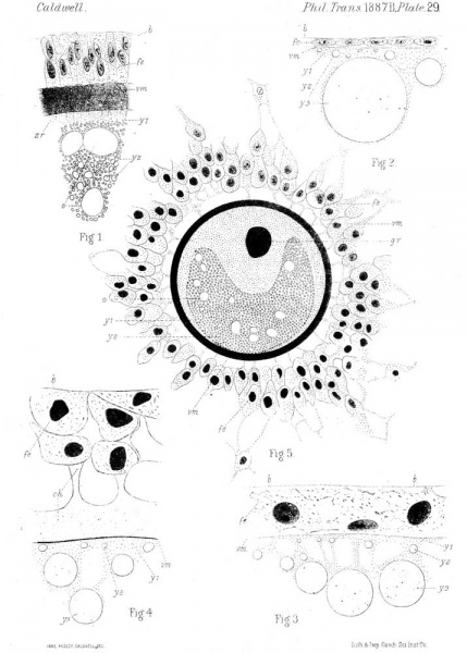
PLATE 29. Sections through Echidna and Phascolarctos
Black and white version of Plate 29.
Fig. 1. Echidna. - Small portion of a section through the ovarian ovum, measuring 0.32 mm. in diameter. 1st period : fe, follicular epithelium ; vm, vitelline membrane ; zr, zona radiata ; i/, yolk granules ; y.., white yolk ; o, oil globule ; b, basilar membrane.
Fig. 2. Echidna. - Small portion of a section through the ovarian ovum, measuring 1 mm. in diameter. 2nd period : lettering as in fig. 1.
Fig. 3. Echidna. - Small portion of a section through the nearly mature ovarian ovum. Beginning of 3rd period : lettering as in fig. 1.
Fig. 4. Echidna. - Small portion of a section through the ripe ovarian ovum, measuring 3 mm. in diameter. 3rd period : eh, pro-albumen.
Fig. 5. Phascolarctos cinereus. - Medium section through a nearly mature ovarian ovum taken from the " liquor folliculi" of a follicle measuring 9 mm. X 6 mm. : fe, follicular epithelium; vm, vitelline membrane ; gv, germinal vesicle.
Zeiss, oo. 2, obj.
Reference Letters
- alb = albumen.
- h = basilar membrane of follicular epithelium
- bl = posterior opening of blastopore.
- ch = pro-albumen.
- ep = epiblast.
- fe = follicular epithelium.
- gv = germinal vesicle.
- hy = hypoblast.
- o = oil (?) globule.
- n1 = nucleus of smaller segmentation area.
- n1 = nucleus of larger segmentation area.
- sh = shell membrane.
- sh1= middle layer of shell membrane.
- sh2= papillae of shell membrane.
- um = coagulum.
- vm = vitelline membrane.
- y1 = yolk granules.
- y2 = white yolk.
- y3= yellow yolk.
- zr = zona radiata.
| Historic Disclaimer - information about historic embryology pages |
|---|
 |
- Links: plate 31 bw | plate 31
Reference
Caldwell WH. The Embryology of Monotremata and Marsupialia Part I. (1887) Phil. Trans. Roy. Soc 178 B.
Cite this page: Hill, M.A. (2024, April 26) Embryology Caldwell01.jpg. Retrieved from https://embryology.med.unsw.edu.au/embryology/index.php/File:Caldwell01.jpg
- © Dr Mark Hill 2024, UNSW Embryology ISBN: 978 0 7334 2609 4 - UNSW CRICOS Provider Code No. 00098G
| Historic Disclaimer - information about historic embryology pages |
|---|
|
File history
Click on a date/time to view the file as it appeared at that time.
| Date/Time | Thumbnail | Dimensions | User | Comment | |
|---|---|---|---|---|---|
| current | 10:51, 31 January 2012 | | 715 × 1,000 (87 KB) | S8600021 (talk | contribs) | {{Caldwell1887}} {{Historic Disclaimer}} |
You cannot overwrite this file.
File usage
The following page uses this file: