File:Brewer1937 plate10.jpg

Original file (1,280 × 1,817 pixels, file size: 323 KB, MIME type: image/jpeg)
Plate 10
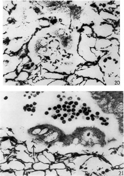
20 The reticulum in the maternal tissue surrminding the multinucleated syncytial cell mass in the center of the picture is irregularly arranged. In many places the strands are clumped, frayed, and sharply broken off. The syncytial mass has phagocytized particles of reticular strands. At one point there is a, phagocytized strand continuous with the reticulum about the syncytial mass. Bielschowsky-Maresch technique. X 790.
21 This photomicrograph shows the junction of the maternal and fetal tissues. The edge of the maternal tissue is indicated by the termination of the reticular framework. The reticulum is frayed and irregular. Numerous strands project freely into the implantation cavity. There is clumping and irregula1'ity of pattern of the reticular frmliework. The syncytial cell mass has phagocytized some reticular strands and continuity between the reticulum and these strands is demonstrated. Bielschowsky-Maresch teclmique. X 790.
| Historic Disclaimer - information about historic embryology pages |
|---|
 |
- Links: plate 1 | fig 1 | fig 2 | fig 3 | plate 2 | fig 4 | plate 3 | fig 5 | fig 6 | plate 4 | fig 7 | fig 8 | fig 9 | fig 10 | plate 5 | plate 6 | plate 7 | plate 8 | plate 9 | plate 10 | plate 11 | plate 12 | plate 13 | plate 14 | fig 11 | 1937 Brewer | Historic Papers | Carnegie Embryo 8819 | Carnegie stage 6 | Week 2
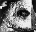
Fig 1 and 2 Tissue block removed
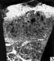
Fig 3 Hemorrhagic region
Fig 4 Blastocyst directly beneath surface hemorrhage
Fig 5 Blastocyst entirely surrounded by short villi
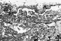
Fig 6 Portion of the chorionic wall shows two short villi
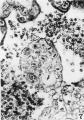
Fig 7 Cytotrophoblastic cell mitosis
Fig 8 Fetal cell is shown amid maternal tissue
Fig 9 Lateral sinus and fetal trophoblast
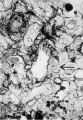
Fig 10 Spiral arteriole with a syncytial cell
Fig 11 Drawing of cytotrophoblastic cells
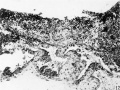
Fig 12 Operculum with one lateral margin
Fig 13 Operculum with adherent villus
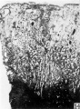
Fig 14 Decidua
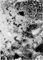
Fig 15 Distal portion of the spiral artery
Fig 16 Spiral artery in the lateral penetration zone
Fig 17 Gland epithelium with syncytial elements
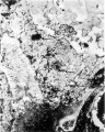
Fig 18 A spiral artery with hemorrhage
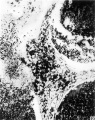
Fig 19 End of this spiral artery in penetration zone
Fig 20 Reticulum in the maternal tissue
Fig 21 Junction of the maternal and fetal tissues
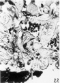
Fig 22 Wall of an endometrial gland
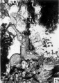
Fig 23 Endometrial gland
Fig 24 Endometrial gland in implantation cavity
Fig 25 Surface epithelium of the decidua
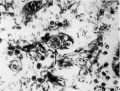
Fig 26 Trophoblastic cell
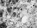
Fig 27 Trophoblastic cell
Fig 28 Trophoblastic cell
Fig 29 Trophoblastic cell phagocytized lymphoyte
Fig 30 Multinucleated syncytial mass


Reference
Brewer JI. A normal human ovum in a stage preceding the primitive streak (The Edwards-Jones-Brewer ovum). (1937) Amer. J Anat., 61: 429-481.
Cite this page: Hill, M.A. (2024, May 15) Embryology Brewer1937 plate10.jpg. Retrieved from https://embryology.med.unsw.edu.au/embryology/index.php/File:Brewer1937_plate10.jpg
- © Dr Mark Hill 2024, UNSW Embryology ISBN: 978 0 7334 2609 4 - UNSW CRICOS Provider Code No. 00098G
File history
Click on a date/time to view the file as it appeared at that time.
| Date/Time | Thumbnail | Dimensions | User | Comment | |
|---|---|---|---|---|---|
| current | 13:15, 3 February 2017 | | 1,280 × 1,817 (323 KB) | Z8600021 (talk | contribs) | |
| 12:00, 3 February 2017 | 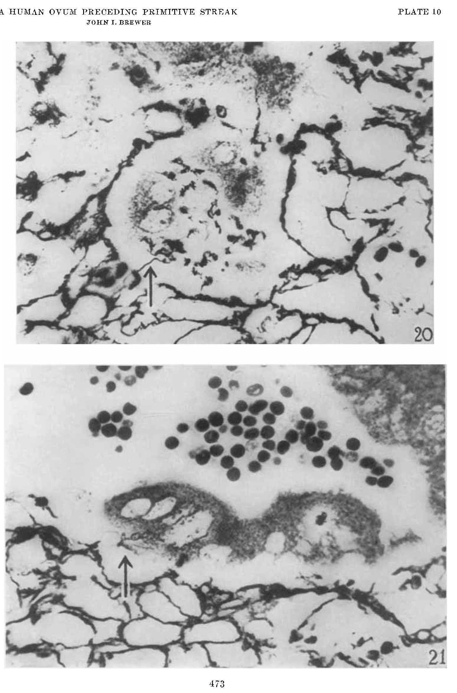 | 1,545 × 2,342 (424 KB) | Z8600021 (talk | contribs) |
You cannot overwrite this file.
File usage
The following page uses this file: