File:Brambell1927a plate31.jpg
From Embryology
Size of this preview: 483 × 599 pixels. Other resolution: 1,739 × 2,157 pixels.
Original file (1,739 × 2,157 pixels, file size: 444 KB, MIME type: image/jpeg)
Plate 31
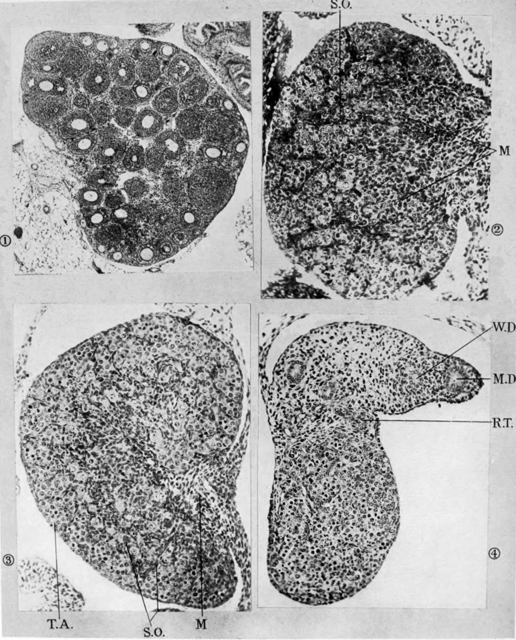
| Fig. 1. Ovary of young mouse three weeks old, showing large number of follicles which have developed precociously and are about to degenerate. X 42. | Fig. 2. 0vary of 17-day embryo showing the formation of the medulla. X 140. |
| Fig. 3. 0vary of new-born mouse showing the structure of the ovary before the growth of the follicles (same fig. as fig. 5 in Plate 7 (4) ). X 140. | Fig. 4. Ovary of 13,1;-day embryo showing many of the germ-cells in synopsis. X 150. |
| Historic Disclaimer - information about historic embryology pages |
|---|
 |
Reference
Brambell FWR. The development and morphology of the gonads of the mouse. Part I. The morphogenesis of the indifferent gonad and of the ovary. (1927) 101: 391-407.
Cite this page: Hill, M.A. (2024, May 3) Embryology Brambell1927a plate31.jpg. Retrieved from https://embryology.med.unsw.edu.au/embryology/index.php/File:Brambell1927a_plate31.jpg
- © Dr Mark Hill 2024, UNSW Embryology ISBN: 978 0 7334 2609 4 - UNSW CRICOS Provider Code No. 00098G
File history
Click on a date/time to view the file as it appeared at that time.
| Date/Time | Thumbnail | Dimensions | User | Comment | |
|---|---|---|---|---|---|
| current | 21:35, 26 April 2018 | | 1,739 × 2,157 (444 KB) | Z8600021 (talk | contribs) | ===Plate 31=== Fig. 1. Ovary of young mouse three weeks old, showing large number of follicles which have developed precociously and are about to degenerate. X 42. Fig. 2. 0vary of 17-day embryo showing the formation of the medulla. X 140. Fig. 3. 0... |
You cannot overwrite this file.
File usage
The following page uses this file: