File:Bradley1908 fig08.jpg
From Embryology
Size of this preview: 707 × 600 pixels. Other resolution: 1,280 × 1,086 pixels.
Original file (1,280 × 1,086 pixels, file size: 229 KB, MIME type: image/jpeg)
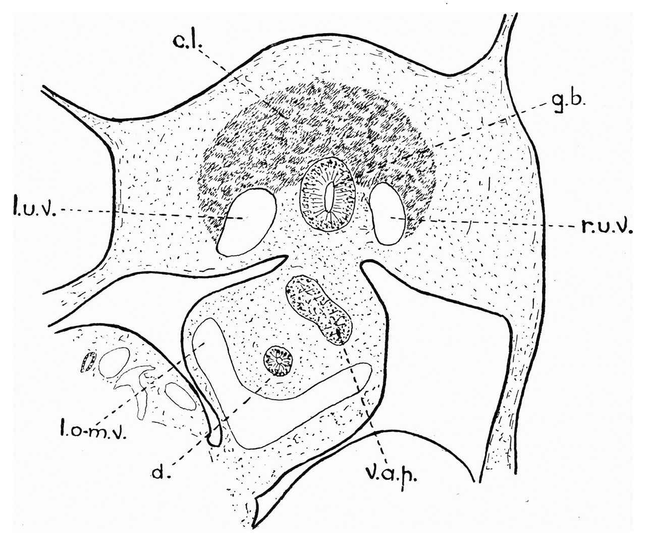
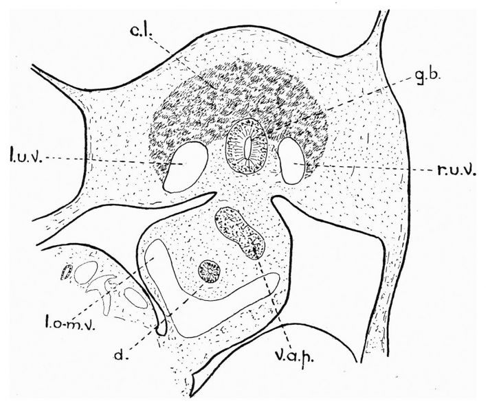
Fig. 8. Camera-lucida drawing of section of liver of 6-mm. pig embryo
v.a.p., ventral anlage of pancreas. The section illustrates the symmetrical disposition of the two umbilical veins, one on each side of the gall-bladder.
Reference
Bradley OC. A contribution to the morphology and development of the mammalian liver. (1908) J Anat. 43: 1-42. PMID 17232788
Cite this page: Hill, M.A. (2024, May 9) Embryology Bradley1908 fig08.jpg. Retrieved from https://embryology.med.unsw.edu.au/embryology/index.php/File:Bradley1908_fig08.jpg
- © Dr Mark Hill 2024, UNSW Embryology ISBN: 978 0 7334 2609 4 - UNSW CRICOS Provider Code No. 00098G
File history
Click on a date/time to view the file as it appeared at that time.
| Date/Time | Thumbnail | Dimensions | User | Comment | |
|---|---|---|---|---|---|
| current | 21:32, 21 November 2019 | | 1,280 × 1,086 (229 KB) | Z8600021 (talk | contribs) | |
| 21:31, 21 November 2019 | 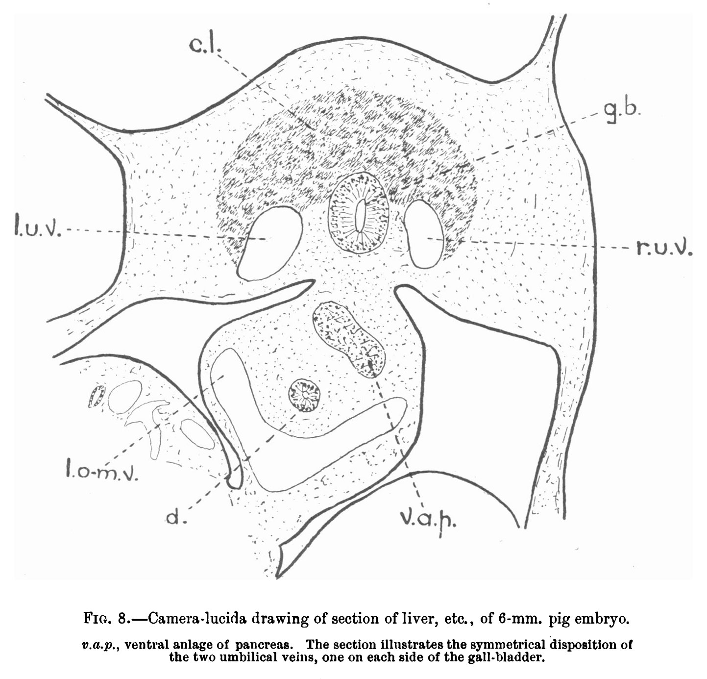 | 1,443 × 1,385 (246 KB) | Z8600021 (talk | contribs) | Fig. 8. — Camera-lucida drawing of section of liver, etc., of 6-mm. pig embryo. v.a.p., ventral anlage of pancreas. The section illustrates the symmetrical disposition of the two umbilical veins, one on each side of the gall-bladder. |
You cannot overwrite this file.
File usage
The following 2 pages use this file: